Whole Slide Scanning and Beyond – MMI CellScan
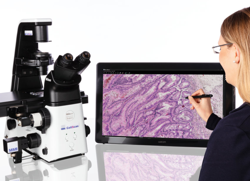
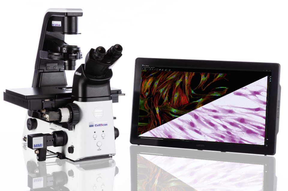
The MMI CellScan is the most flexible whole slide imaging system on the market. With compatibility across a wide range of research microscopes and any possible imaging mode, CellScan maximizes the performance of your research or pathology services.
CellScan is a whole slide scanner that converts any sample format into high-resolution digital data in 5D: Time-lapse, Z-stack, XY Scan, and multi-color fluorescence for the deepest insights into your sample.
CellScan is not just a whole slide scanner; it’s a comprehensive imaging solution tailored to meet the demands of modern research. Elevate your imaging experience with CellScan’s advanced features and take your research to new heights.
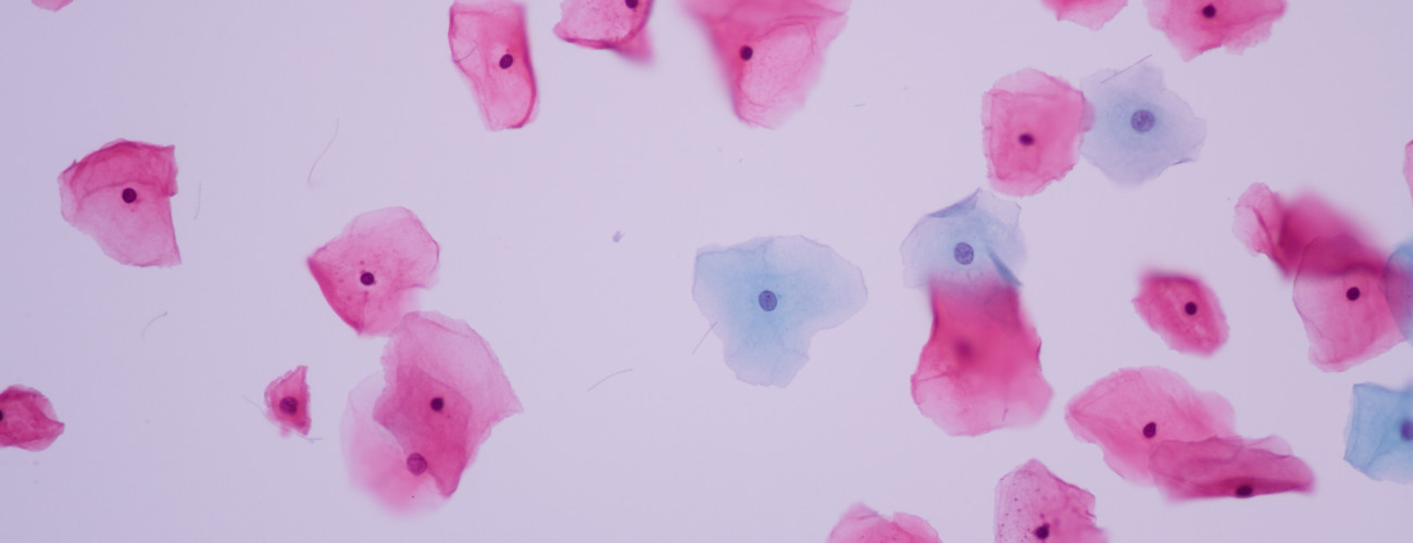
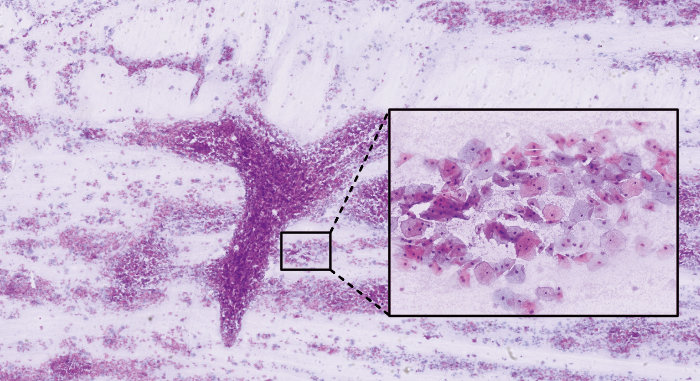
Epithelial cells of cervical human – 40x objective
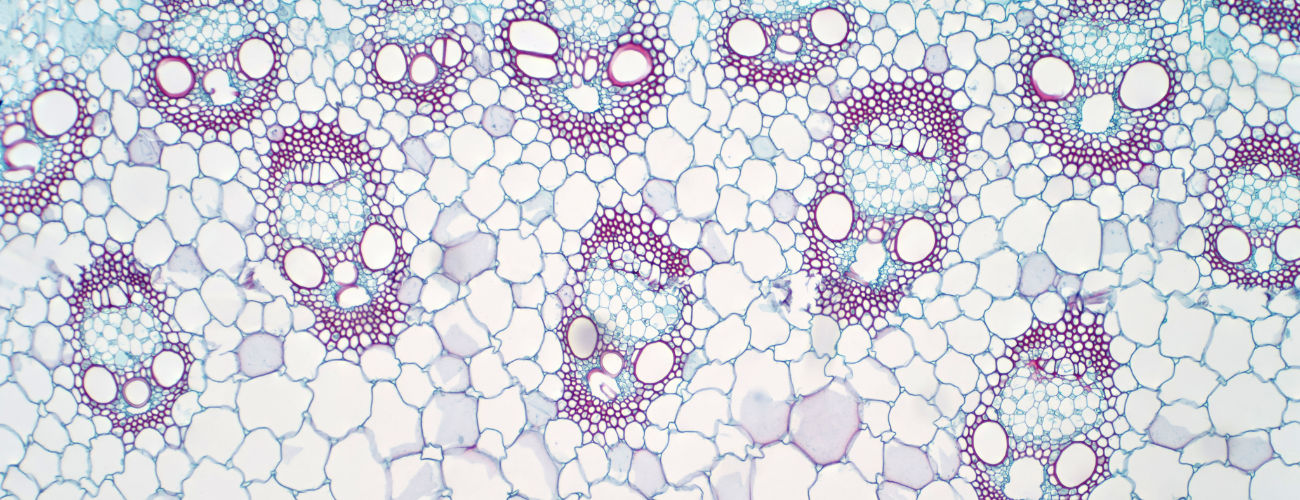
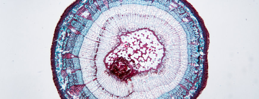
Monocot plant vascular tissue – 20x objective
What is a whole slide scanner?
Whole slide imaging (WSI) – also known as virtual microscopy or whole slide scanning – is becoming increasingly popular in digital pathology as well as in clinical and basic research. A whole slide scanner can take images of each field of view across an entire microscopy slide. The pictures are then stitched together to generate a single, high-resolution digital whole slide image that researchers use for various purposes.
Why whole-slide imaging?
Whole slide imaging offers many advantages over conventional microscopy, such as portability, ease of sharing, retrieval of images, workload balancing, and image analysis. Scientists use whole slide scanners in many clinical and nonclinical settings. Clinical scientists are particularly excited about its use in digital pathology, where published data shows an excellent correlation between diagnoses made with whole slide imaging and conventional light microscopy. Moreover, biotech and pharmaceutical companies use whole slide scanners to reduce costs, and improve standardization and data management in research and development.
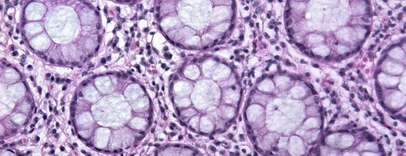
Glandular tissue – 20x objective
Tilia Stem – 40x objective
“The MMI CellScan whole slide scanner supports us in our daily work: this tool documents our tissue sections in high resolution and it fully integrates into our laser microdissection workflow. We especially appreciate that we can annotate directly in the image thus saving hands-on time at the instrument.”
The most flexible whole slide scanner
Compatible with almost all microscopes and imaging modes
The MMI CellScan is compatible with almost all research microscopes. You decide what microscope to use. Therefore, CellScan is not just a whole slide scanner, it’s a gateway to limitless digital imaging possibilities. Whether you’re working with brightfield, multi-fluorescence, confocal, phase contrast, or any other imaging mode, this versatile whole slide scanner gives you the freedom to explore and capture your samples in unprecedented detail.
 CellScan supports all image modes
All common sample formats
The whole slide scanner CellScan supports various sample formats. From conventional glass slides (including coverslips) to petri dishes with living cells, and membrane slides designed for laser microdissection. This unmatched flexibility ensures that CellScan is tailored to meet the diverse needs of various applications, like in digital pathology, cell biology and oncology.
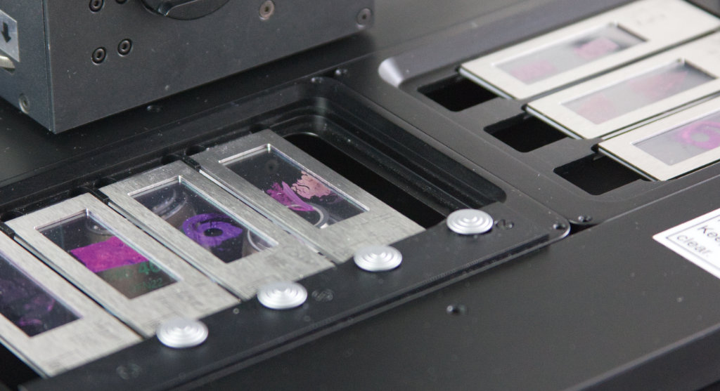 Open x-y stage with four membrane slides
5D imaging
Take control of your research with CellScan’s ability to scan full-resolution digital slides in 5D. Capture dynamic changes over time with time-lapse imaging, explore the depths of your samples in Z-stack mode, navigate seamlessly in the x-y axis, and unravel the complexity of multi-fluorescence imaging. The MMI whole slide scanner empowers you to conduct experiments and analyse exactly as you envision.
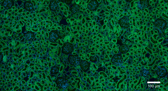 Rat kidney with multi-fluorescence (FITC and DAPI)
Modularity is key
The whole slide scanner CellScan boasts exceptional modularity, offering seamless compatibility and upgradability with all MMI devices. With modules like CellCut for precise laser microdissection, CellEctor for isolating cells in suspensions, and CellDetector utilizing artificial intelligence for automatic cell and tissue identification, our whole slide scanner provides a versatile platform for evolving research needs.
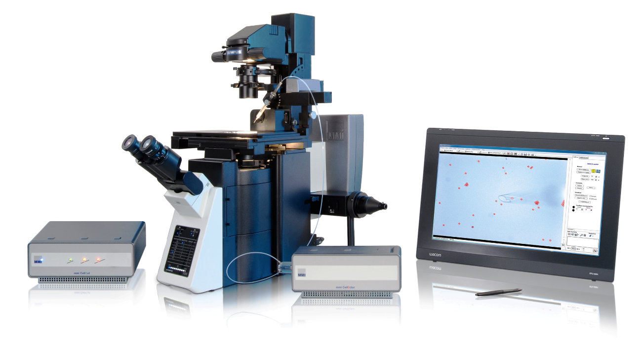 CellScan with the CellCut and CellEctor on the Olympus IX83
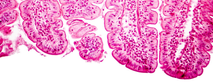
Villi of small intestine – 100x objective
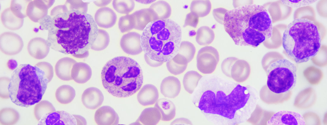
White blood cells in a blood smear – 100x objective
CellScan in a nutshell
It’s a whole slide scanner
- Full resolution in any magnification
- Any sample type
- In 5D (x, y, z, time, colour)
- Open file format BigTIFF
It’s a microscope
- Anything you would do with a microscope (e.g. any objective lenses and fluorescence filter)
- Any image mode, including confocal
Its upgradeable
- With various microscope accessories
- With any other MMI system (laser microdissection, cell picking and automated cell/tissue detection)
MMI imaging Software at the forefront of innovation
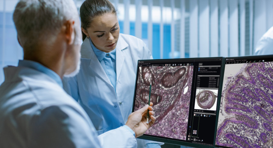
Collaborate with ease
Empower your whole slide imaging experience with precision and ease. The MMI analyzing software comes with unlimited licenses, facilitating remote and interactive work within larger teams. The whole slide images are loaded instantaneously as BigTIFF. The open file-format allows you to open the images in a range of other image analysis software.
 Access samples from anywhere at anytime
Capture and annotate
Mark and annotate cells and regions of interest effortlessly, with the added capability to export and reload marked areas. Insert scale bars, measure distances, and utilize this functionality for laser microdissection with the MMI CellCut. Capture videos and images at any time, providing flexibility in documentation.
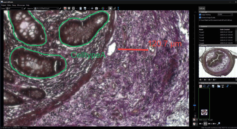 Make annotations anywhere
Stitching perfection for best image harmony
The MMI whole slide scanner boasts a sophisticated stitching function, ensuring a perfect alignment of individual images without any overlap or artefacts along the edges. This precision is achieved through the accuracy of the motorized stage, offering absolute positioning and sample localization. The stability of the microscope set-up, coupled with precise auto-focusing, particularly benefits samples with waviness along the z and focus directions.
 Vaginal swab recorded by CellScan
If you have any questions about the CellEctor cell picker, please feel free to contact us
Contact us nowWhole slide live-cell imaging
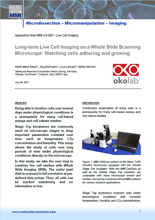
Live cell imaging involves time-lapse microscopy on living cells. Scientists use it to better understand biological function and cellular dynamics. With the whole slide scanner CellScan in combination with a stage-top incubator, you can easily perform live cell imaging experiments in fully physiological cell culture conditions.
Read more in the following Application Notes:
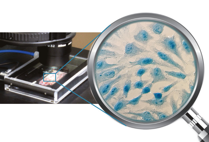 HeLa cells stained with Coomassie blue
Capturing and compiling multi-colour fluorescence images
CellScan is a whole slide imaging system that can utilize all functions of a research microscope. Therefore, the scanning of whole slides is not limited to one resolution. Researchers can change objectives or switch between brightfield and fluorescence imaging modes with just one mouse click. You can collect digital images of single fluorescent channels and automatically superpose them to generate one multi-colour image file.
Read more in the following Application Note:
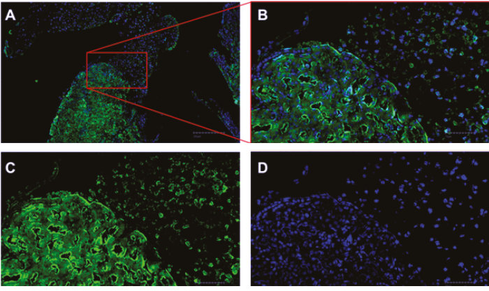 A) Overview image. B) Both channels DAPI (blue) and CF 594 (green). C) CF 594. D) DAPI
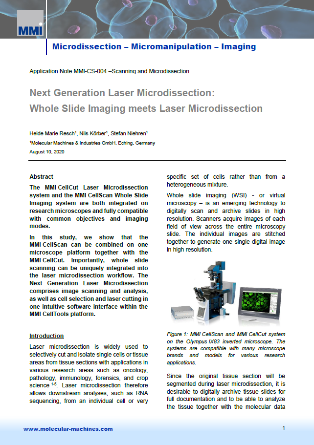
Whole slide imaging meets laser microdissection
Researchers from a multitude of fields use microdissection to selectively isolate single cells or tissues. The technique enables downstream analyses such as RNA sequencing. Scientists often discard the larger cut tissue section, losing valuable information from the original slide. However, MMI’s CellScan, in combination with the CellCut Laser Microdissection system, allows researchers to image a tissue section and then precisely select cells and dissect the tissue while preserving information about the original uncut tissue and position information about the cut.
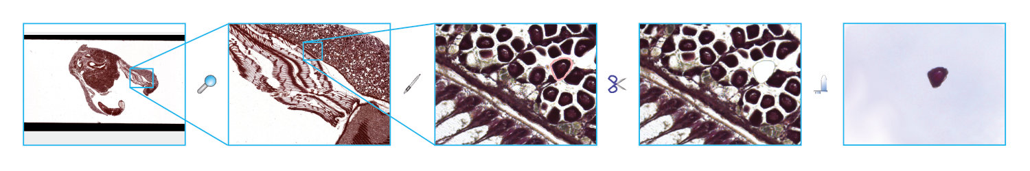
1. Scan your slide 2. Identify target region 3. Mark target cell 4. Excise the target cell 5. Verify successful isolation
Have a look at how the whole slide scanner works
In the following video you will see how to scan a slide with CellScan and how to cut out a cell with the optional laser microdissection module CellCut.

By loading the video, you agree to YouTube’s privacy policy.
Learn more
MMI CellScan whole slide scanner: Application Notes
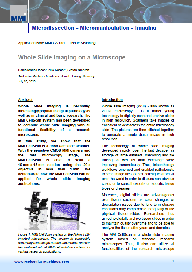
Tissue Scanning
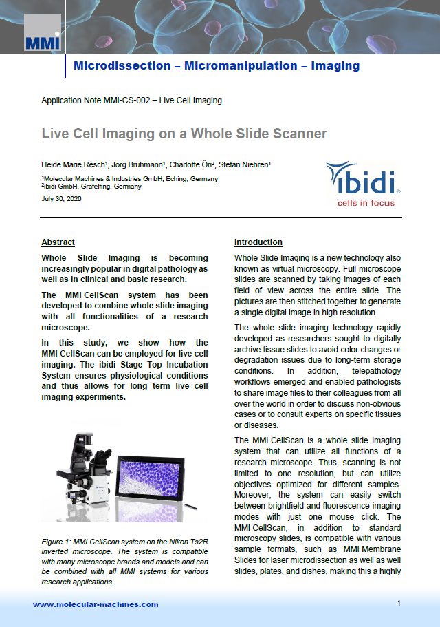
Live Cell Imaging
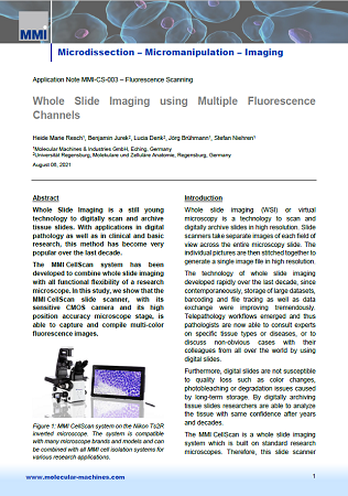
Fluorescence Scanning
Whole Slide Imaging meets Laser Microdissection
Long-term Live Cell Imaging on a Whole Slide Scanner
MMI CellScan whole slide scanner: Documents
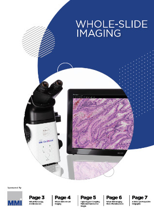
White Paper
Whole Slide Imaging and Beyond
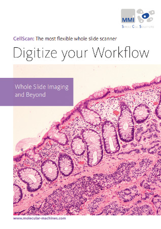
Flyer
Most versatile WSI system
User Manual
How can we support your project?