
Laser Capture Microdissection
Short definition:
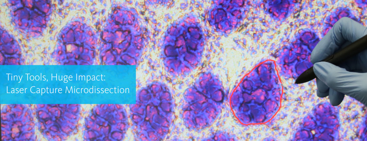
Laser Capture Microdissection is defined as the precise cutting of microscopic tissue area using a laser and the subsequent separation of the dissected area from surrounding tissue. Importantly, laser dissection microscopes differ in the particular method to isolate target cells after cutting. MMI for example isolates cut tissue by employing adhesive isolation caps, which gently and reliably take up the sample of any size and shape.
What is laser capture microdissection?
Laser Capture Microdissection is defined as the precise cutting of microscopic tissue area using a laser, and the subsequent separation of the dissected area from surrounding tissue. Terms such as Laser Microdissection, Laser-assisted Microdissection, Microdissection, Laser Capture or Laser Dissection are used synonymously and describe the same method. The first commercial laser microdissection microscopes have been developed and built in the late 1990s to early 2000s, based on the research by Michael R. Emmert-Buck and his colleagues at the National Institutes of Health and the National Cancer Institute (Bethesda, Maryland, USA). The following years, the technology has been further improved to allow for more flexible and automated workflows, for easier handling as well as for more reliable and precise cell isolation. Therefore, also the isolation of single cells from samples such as tissue sections, smears or live cell culture is now possible. Intriguingly, single cell sequencing technologies have emerged over the last decade which now enable molecular analyses of single cells isolated by laser capture microdissection. Single cell transcriptomics or proteomics methods have been established and improved to shed light on various molecular mechanisms in individual cells.Your benefits using laser microdissection
Have a look how it works
This video shows a typical laser microdissection workflow:
By loading the video, you agree to YouTube's privacy policy.
Learn more
How does laser capture microdissection work?
Typically, laser capture microdissection systems are based on research microscopes. Lasers are required to precisely cut tissue on this microscopic scale, and they are coupled into the optical paths of the dissection microscopes. For cutting, the sample is mounted on membrane slides to enable both for precise cutting and for subsequent cell isolation procedures. The laser microdissection microscopes on the market today mainly differ in the laser types they use and how the laser is applied to cut the sample. The MMI CellCut uses a low-damage laser with low power but high pulse frequency to allow for efficient and precise cutting and to not compromise tissue integrity. Moreover, the laser is fixed during the cutting process to ensure that the laser is always in focus and the sample is efficiently cut. Importantly, the laser dissection microscopes differ in the particular method to isolate target cells after cutting. MMI for example isolates cut tissue by employing adhesive isolation caps, which gently and reliably take up the sample of any size and shape.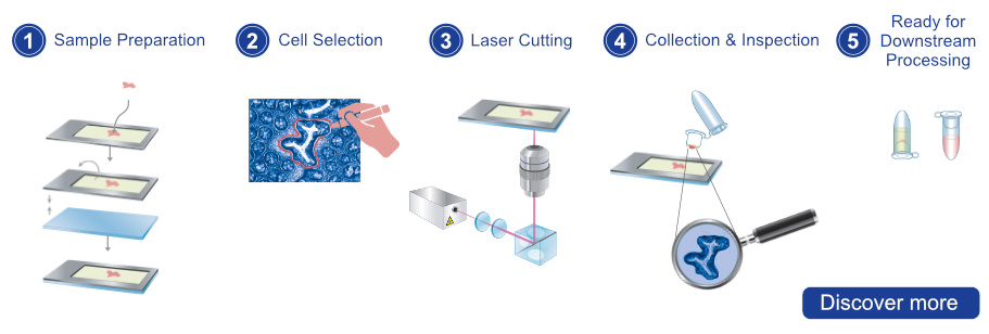
1. Sample Preparation: The tissue sample is first prepared for microdissection by fixing, embedding, and sectioning it into a membrane slide that is inert and has negligible autofluorescence. Afterwards, the membrane slide is inverted and placed onto a glass slide for protection against contamination.
3. Laser Cutting: Once the region/cells of interest have been identified, the laser is focused on the selected area. The thin laser cutting path enables a precise and gentle extraction at an outstanding speed.
4. Collection & Inspection: The isolated target cell is collected by an adhesive cap. The orientation and morphology will be maintained. After cutting the cell can be visualized on the cap.
5. Ready for Downstream Processing: After microdissection, the collected cells can be processed further for any downstream application (e.g. gene expression analysis, RNA isolation and much more)