
New Publication: Expression of G-Protein-Coupled Estrogen Receptor (GPER) in Whole Testicular Tissue and Laser-Capture Microdissected Testicular Compartments of Men with Normal and Aberrant Spermatogenesis
Infertility is a problem that affects approximately 15% of couples, and the male factor is believed to contribute alone or in combination with female factor to about 50% of the cases.
This study is the first one to report increased expression of GPER in testicular tissue of men with primary testicular failure. An application of the laser-capture microdissection technique and the analysis of GPER transcriptional level in separate testicular compartments allowed us to unequivocally demonstrate altered overexpression of GPER mRNA in Sertoli, but not in Leydig cells, which was confirmed at the protein level.
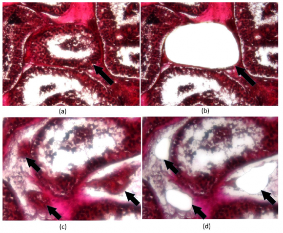
To demonstrate the specificity of this finding, an analysis of contamination with other cell types of our laser-microdissected compartments (i.e., seminiferous tubules and Leydig cell clusters) was conducted and revealed marginal expression of mRNA corresponding to genes strongly and specifically expressed in cell types of seminiferous tubules and interstitium. The mean transcriptional expression of the Sertoli cell marker (FSHR) was very low in laser-microdissected Leydig cell clusters compared with the expression observed in laser-microdissected seminiferous tubules with complete spermatogenesis (Supplementary Materials, Figure S4a). The latter were barely contaminated with CYP17A1 expression in comparison with its expression in laser-microdissected Leydig cell clusters (Supplementary Materials, Figure S5a). Additionally, laser-microdissected Leydig cell clusters demonstrated weak or no transcriptional expression of EMR1 (macrophage marker), CMA1 (mast cell marker) and ACTA2 (peritubular cell marker); their mean expression was comparable to that of FSHR (Supplementary Materials, Figure S4b). Similarly, the level of ACTA2 in laser-microdissected seminiferous tubules was marginal and comparable to the one observed for CYP17A1 (Supplementary Materials, Figure S5b).
Furthermore, the authors also showed that this increased GPER expression in Sertoli cells was related to elevated FSH levels, as well as increased expression of AMH transcripts. Whether the observed change in GPER expression in the present study is a marker of Sertoli cells maturational state, or whether it is involved in the pathogenesis of observed disturbances, needs to be further elucidated.
Read the full publication for further information how to isolate RNA in a contamination-free way for RNAseq analysis.