Whole Slide Scanning and Beyond – MMI CellScan
The MMI CellScan is the most flexible whole slide imaging system on the market. With compatibility across a wide range of research microscopes and any possible imaging mode, CellScan maximizes the performance of your research or pathology services.
CellScan is a whole slide scanner that converts any sample format into high-resolution digital data in 5D: Time-lapse, Z-stack, XY Scan, and multi-color fluorescence for the deepest insights into your sample.
CellScan is not just a whole slide scanner; it’s a comprehensive imaging solution tailored to meet the demands of modern research. Elevate your imaging experience with CellScan’s advanced features and take your research to new heights.
What is a whole slide scanner?
Whole slide imaging (WSI) – also known as virtual microscopy or whole slide scanning – is becoming increasingly popular in digital pathology as well as in clinical and basic research. A whole slide scanner can take images of each field of view across an entire microscopy slide. The pictures are then stitched together to generate a single, high-resolution digital whole slide image that researchers use for various purposes.
“The MMI CellScan whole slide scanner supports us in our daily work: this tool documents our tissue sections in high resolution and it fully integrates into our laser microdissection workflow. We especially appreciate that we can annotate directly in the image thus saving hands-on time at the instrument.”
CellScan in a nutshell
It’s a whole slide scanner
- Full resolution in any magnification
- Any sample type
- In 5D (x, y, z, time, colour)
- Open file format BigTIFF
It’s a microscope
- Anything you would do with a microscope (e.g. any objective lenses and fluorescence filter)
- Any image mode, including confocal
Its upgradeable
- With various microscope accessories
- With any other MMI system (laser microdissection, cell picking and automated cell/tissue detection)
If you have any questions about the CellEctor cell picker, please feel free to contact us
Contact us nowWhole slide imaging meets laser microdissection
Researchers from a multitude of fields use microdissection to selectively isolate single cells or tissues. The technique enables downstream analyses such as RNA sequencing. Scientists often discard the larger cut tissue section, losing valuable information from the original slide. However, MMI’s CellScan, in combination with the CellCut Laser Microdissection system, allows researchers to image a tissue section and then precisely select cells and dissect the tissue while preserving information about the original uncut tissue and position information about the cut.

1. Scan your slide 2. Identify target region 3. Mark target cell 4. Excise the target cell 5. Verify successful isolation
Have a look at how the whole slide scanner works
In the following video you will see how to scan a slide with CellScan and how to cut out a cell with the optional laser microdissection module CellCut.

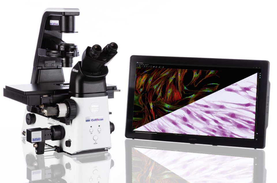 CellScan supports all image modes
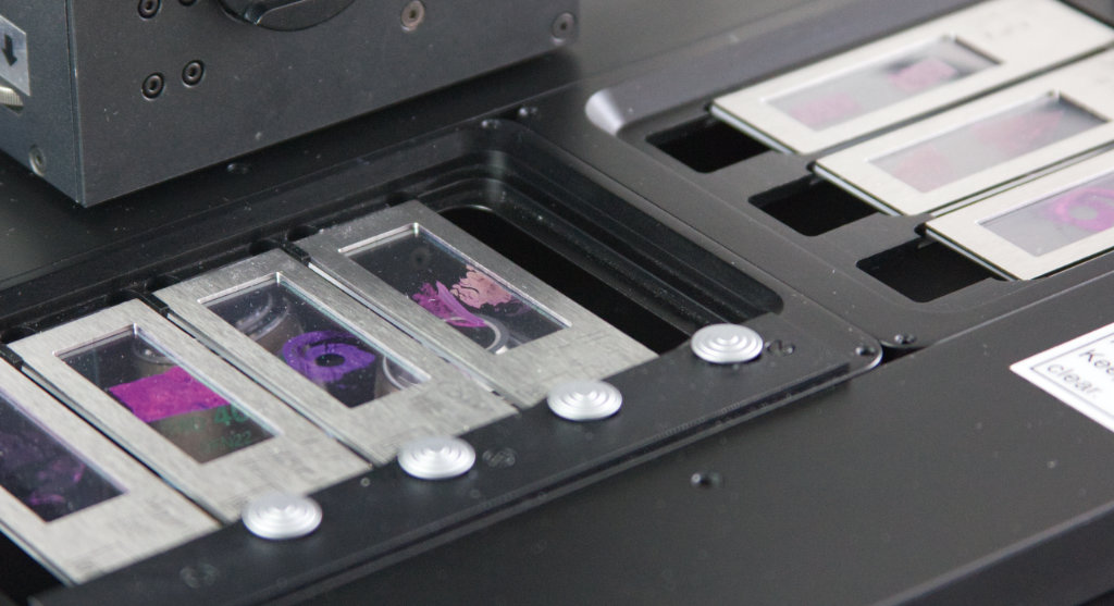 Open x-y stage with four membrane slides
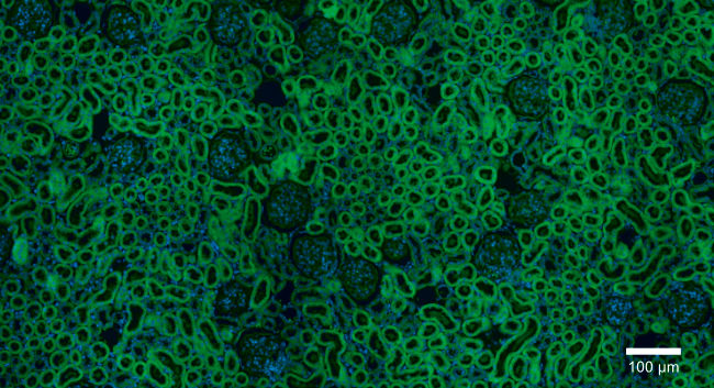 Rat kidney with multi-fluorescence (FITC and DAPI)
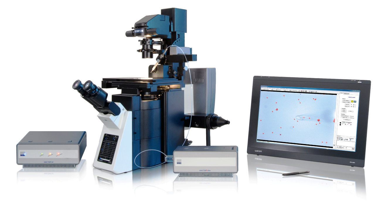 CellScan with the CellCut and CellEctor on the Olympus IX83
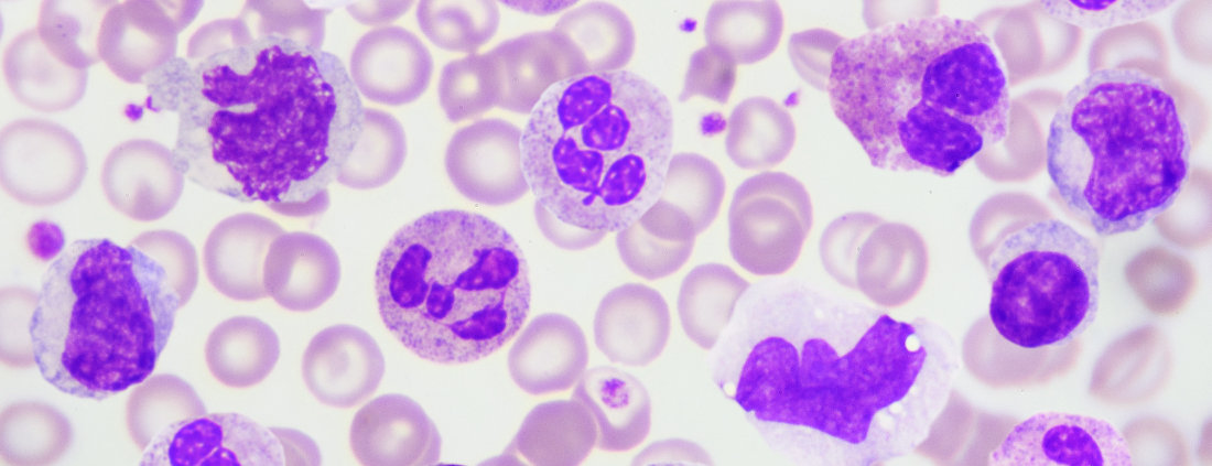
 Access samples from anywhere at anytime
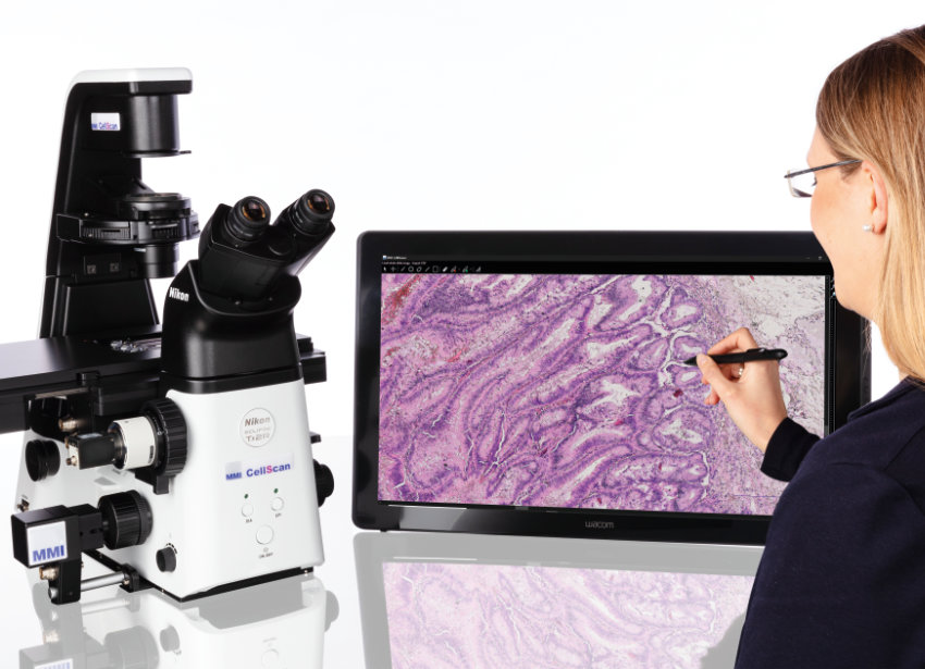
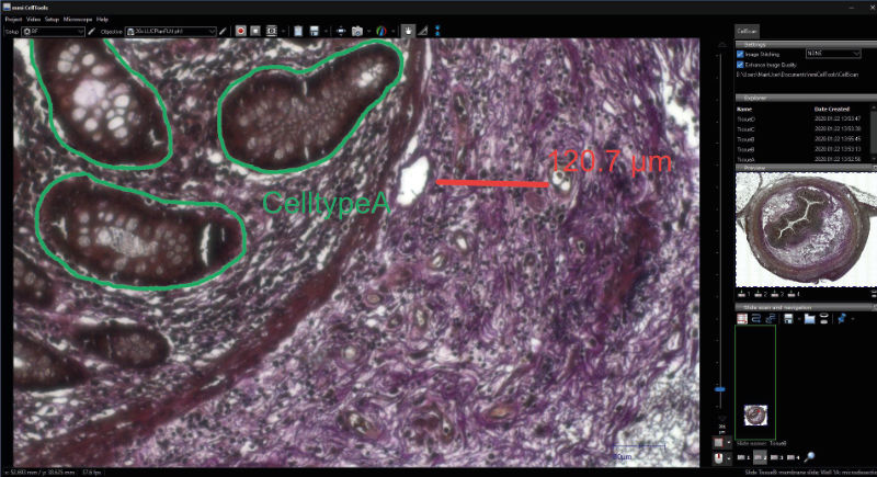 Make annotations anywhere
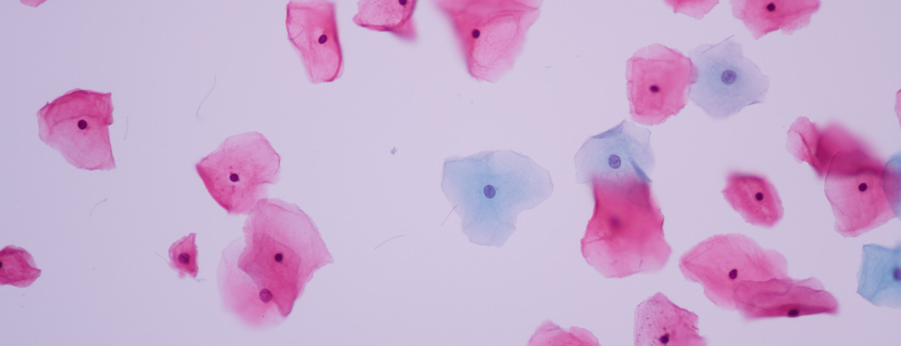
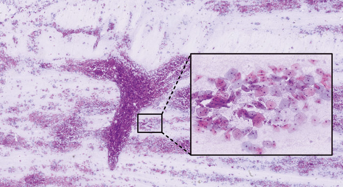 Vaginal swab recorded by CellScan
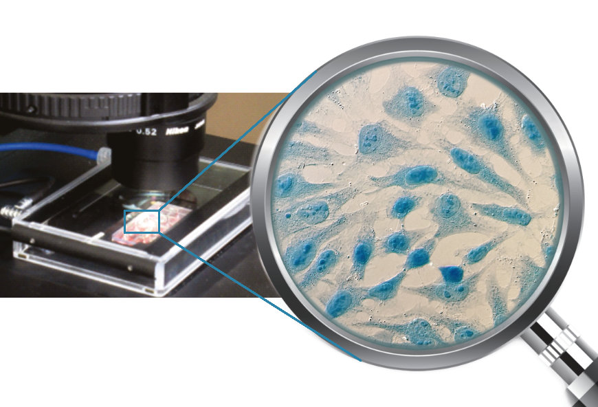 HeLa cells stained with Coomassie blue
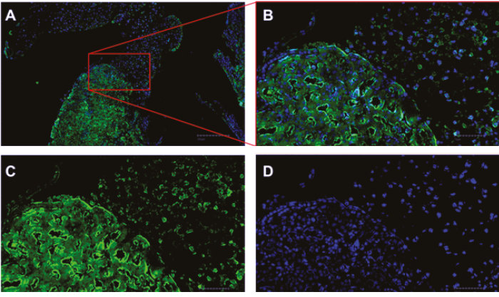 A) Overview image. B) Both channels DAPI (blue) and CF 594 (green). C) CF 594. D) DAPI
