


Manipulate single cells and sub-cellular structures with precision
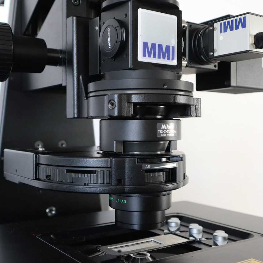
The MMI CellManipulator optical tweezers provide a precise approach to manipulating particles at the microscopic level, enabling researchers to non-invasively handle single cells and delicate sub-cellular structures with exceptional control. By allowing fully contact-free positioning and movement, the CellManipulator optical tweezers preserve the integrity and viability of even the most sensitive samples. This combination of precision and ease of use not only enhances experimental reliability, but also accelerates discoveries in cell biology, molecular research, and a wide range of biomedical applications.
Keep it Flexible
CellManipulator: The optical tweezers system designed for maximum flexibility. Compatible with almost all inverted or upright research microscopes. It can be integrated with fluorescence, TIRF, confocal microscopy, and many more customized adaptations.
Huge Work Space
The entire laser beam control of the optical tweezers is located underneath the trapping lens. This open-space architecture provides unrivalled free space above the optical trap. Perfect for customizable force measurements and other applications.
Become an Expert in 10 min
With the intuitive and flexible MMI CellTools analysis software, you can simply organize and manipulate your optical traps. Optical trapping was never so easy.
Engineered for Versatile Use, with Unyielding Trap Power
Trapped particles cannot escape the experiments inside a petri dish, microfluidic chamber, or simply on a glass surface using the optical tweezers. Never lose your particle with 1200 pN of escape force.
Engineered for Precise & Reliable Force Measurements
Tailor your force spectroscopy setup to your needs—choose imaging-based detection for larger sample workspaces or forward interferometric position sensing for sub-piconewtons force resolution.
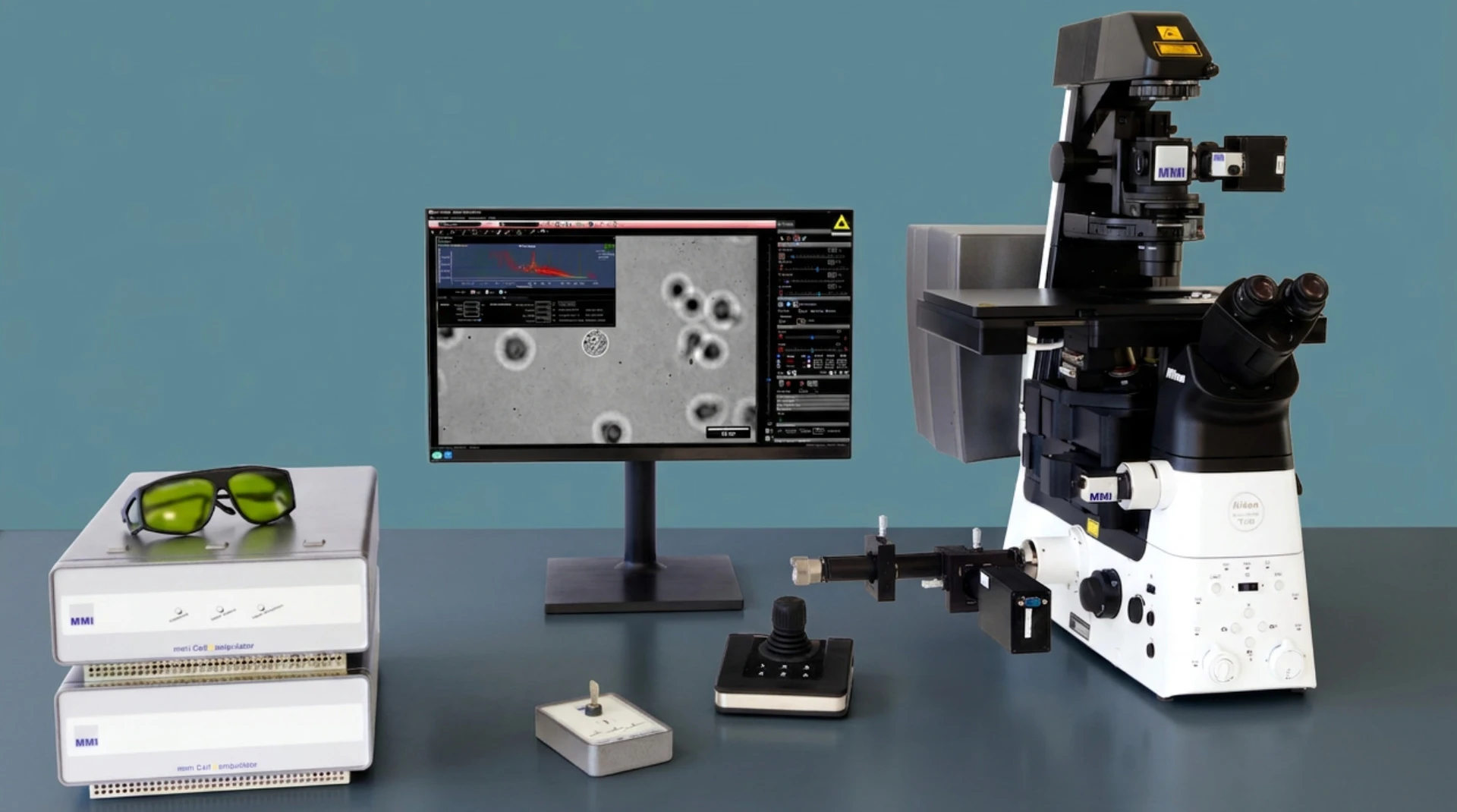
Software
User-friendly CellTools software for data acquisition, visualisation, and interpretation.
Imaging Mode
Imaging-based force spectroscopy. Automatic optical trap stiffness calibration with no compromise in the sample workspace.
Multi-Trap
Time shared up to 20 particles (Galvo steered)
Time shared up to 200 particles (AOM steered)
Back-Focal Plane Mode
Forward Interferometric position detection-based force spectroscopy. Automatic optical trap stiffness calibration with sub-piconewton resolution.
Flexibility
Compatible with leading inverted or upright research microscopes
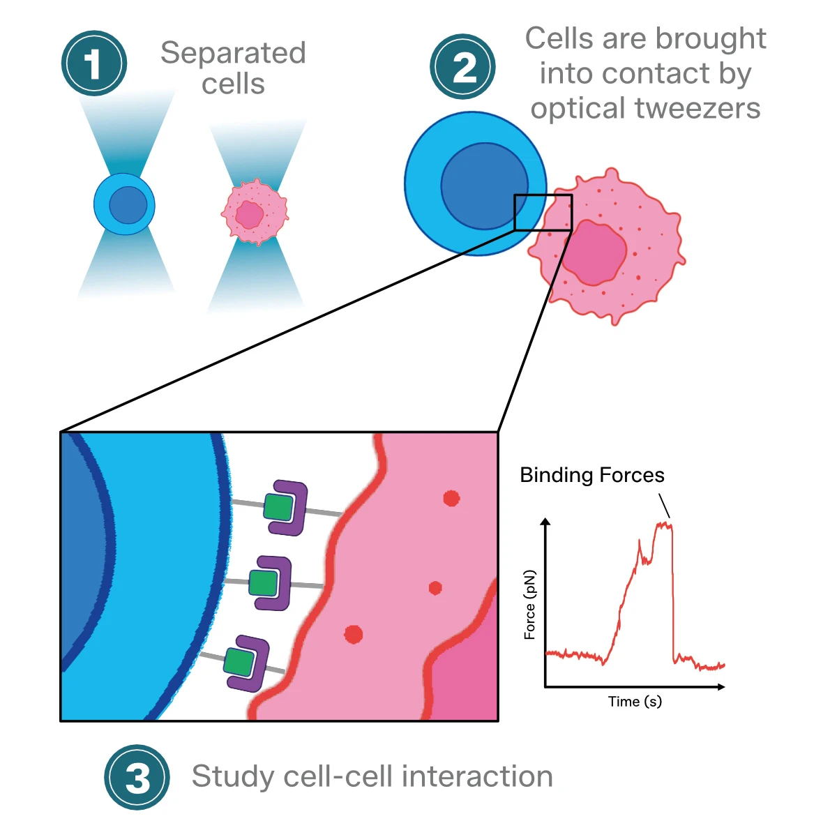
The MMI optical tweezers enable the manipulation of cells to promote cell-cell interactions and directly measure their binding forces while maintaining complete control over the cell’s orientation and the time of the cell-cell contact. This is incredibly valuable for conducting dynamic studies on cell-cell interaction forces and measuring cell avidity with single-cell resolution.
Intracellular Manipulation
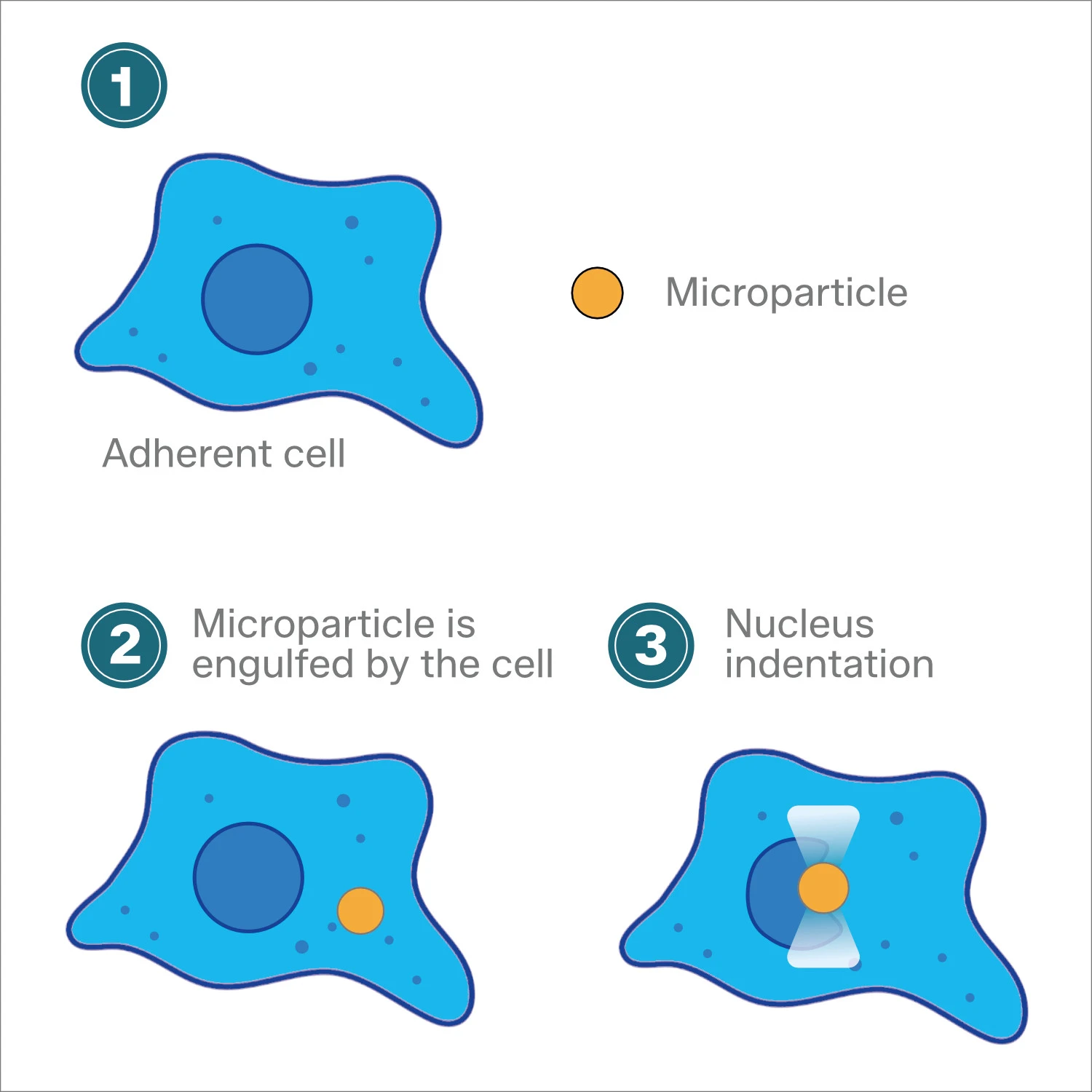
Another common application of optical tweezers is to study the role of the nucleus in mechanotransduction by measuring the force and material properties that shape the cell nucleus inside living cells. Internalised microparticles, like lipid droplets, enable the stretching and indenting of the cell nucleus while monitoring time-force and time-displacement curves. Therefore, with the MMI CellManipulator optical tweezers, it becomes feasible to gather information about viscoelastic properties and stiffness of the cell nucleus.
Measuring nucleus mechanics
Molecular Motor Studies
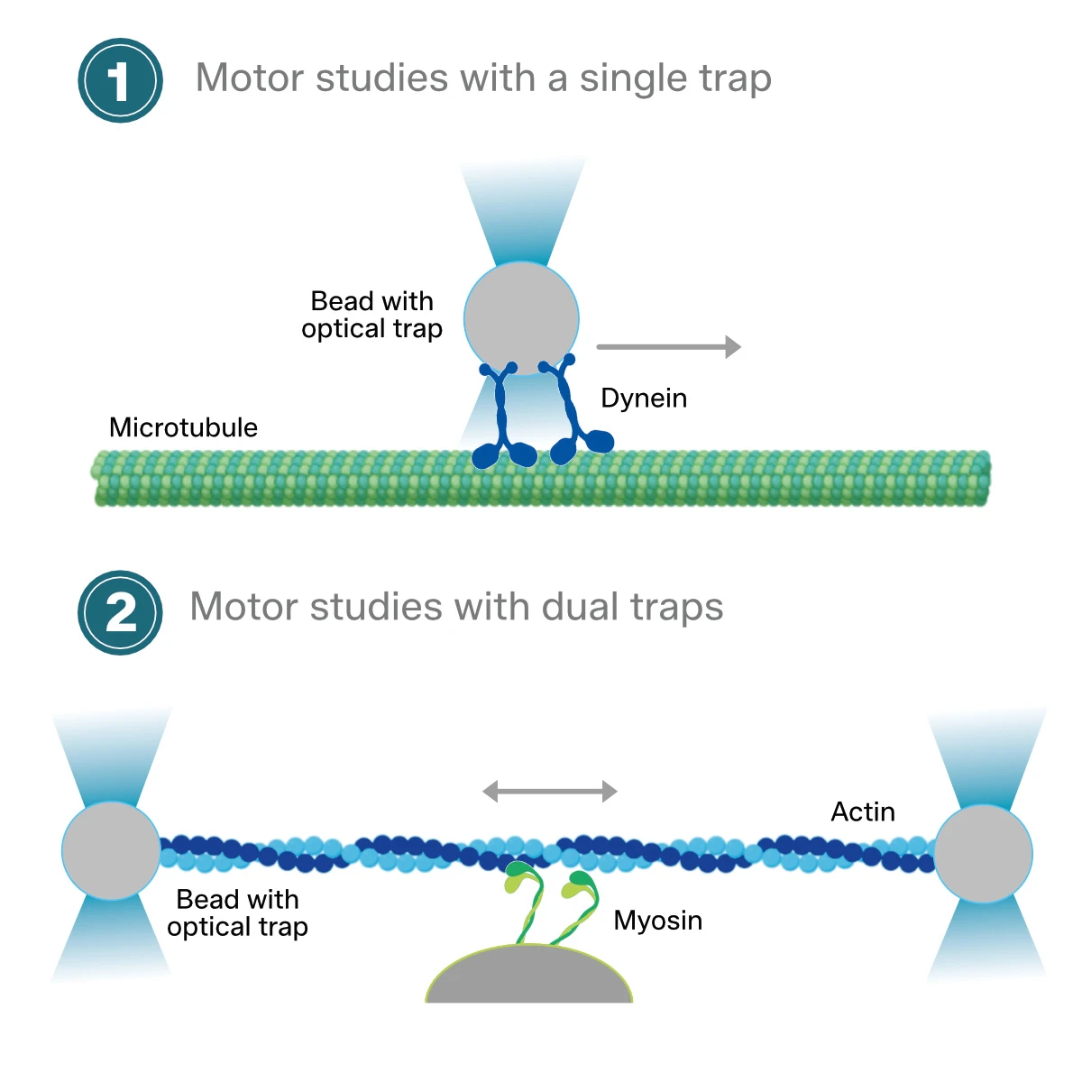
Thanks to numerous technical improvements, optical trapping with the MMI CellManipulator allows synthesizing up to 10 multiple independent optical traps and to measure the forces of motor proteins in sub-pN resolution.
In optical tweezers experiments, the molecule of interest needs to be attached at two points: one end to a small microparticle, and the other end to a fixed substrate or a second microparticle in a separate optical trap. The following visualization shows two typical applications to study motor proteins:
Measuring motor protein sub-pN forces
Laser Tweezers Raman Spectroscopy
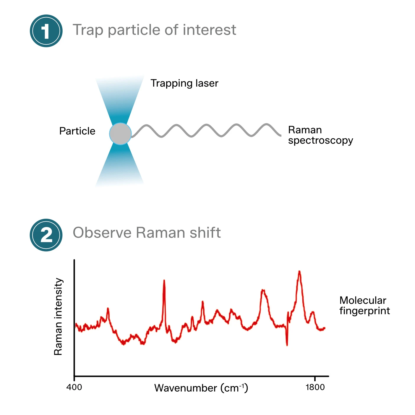
Raman spectroscopy is an analytical technique, based on the inelastic scattering of photons upon interacting with matter. The inelastic scattered photons have different wavelengths/colours compared to the incoming photons generated from the light source. The spectrum generated is dependent on the chemistry of the object. Raman spectroscopy in life science is used to perform so-called “molecular fingerprinting”. Each protein has a unique biochemistry and therefore generates a unique Raman spectrum. The combination of the MMI optical tweezers with Raman spectroscopy can be applied to detect the upregulation of specific proteins in cancer cells.
Obtain the molecular fingerprint
Isolation and Manipulation of Rare Cells
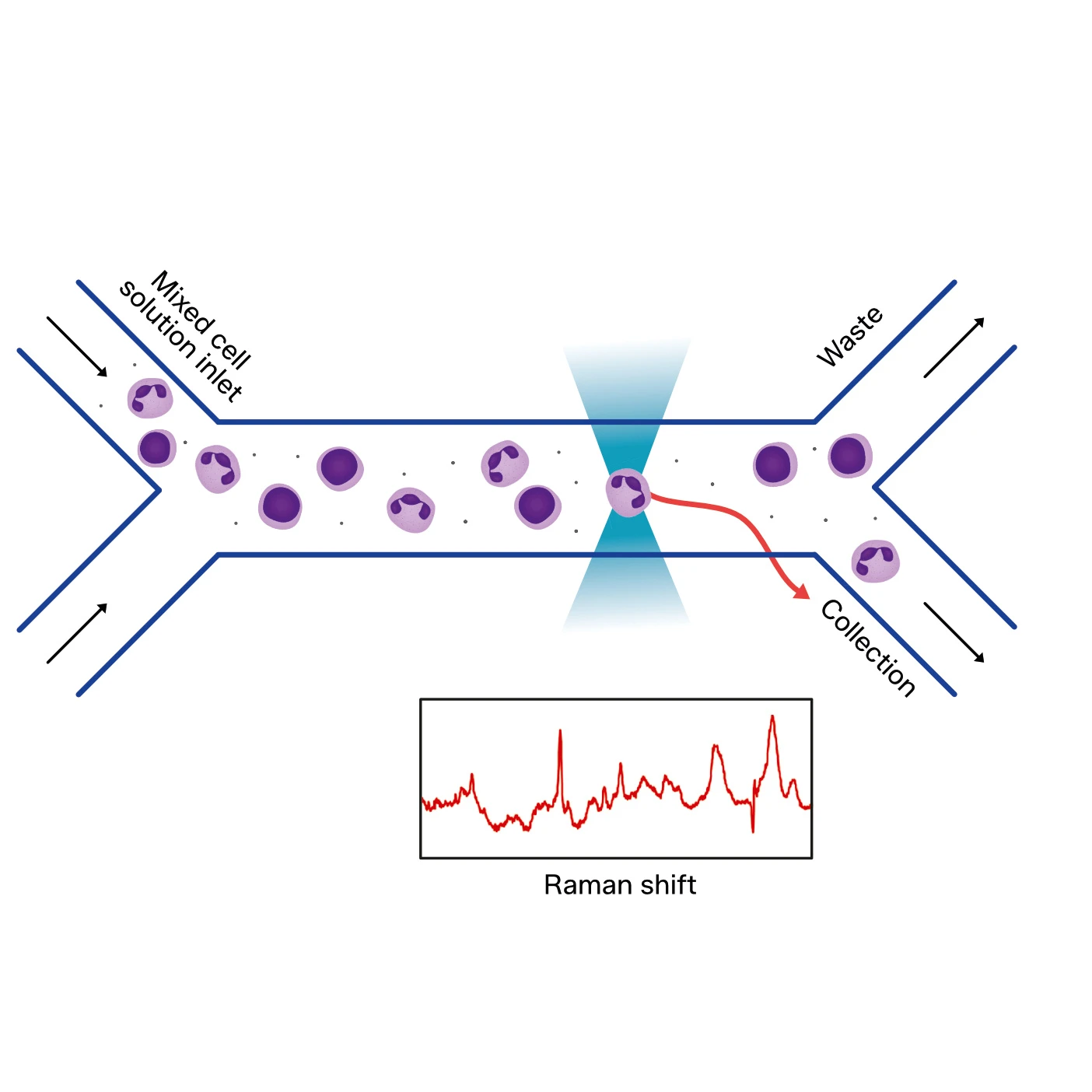
Combining optical tweezers and microfluidic chip technologies enables the isolation and manipulation of rare cells with high recovery and purity, which is crucial for downstream multi-omics profiling.
The combination of the MMI CellManipulator optical tweezers with microfluidic chip technology enables the real-time sorting of small cell populations with high accuracy. The systemstarts by using a laminar flow to focus the targeted cells. The optical tweezers can then precisely move the cell in a non-invasive manner to the desired destination. In contrast to traditional cell sorting technologies, such as flow cytometry, cell sorting with optical tweezers has the unique advantage of its high recovery rate, flexibility, and purity in small cell population sorting.
Cell Sorting with optical tweezers

Universal Laser
Piezo Stage Minimum Step Size = 0.01 um (lateral)
Piezo Stage Field of Movement = 0.1 x 0.1 mm
Motorized Stage Field of Movement = 120 x 80 mm
Time-gated autofluorescence microscopy of motile green microalga in an optical trap: Koenig et al. (1998). Cell Mol Biol.
https://pubmed.ncbi.nlm.nih.gov/9764746/
Cell sorting: Divide and conquer: Michael Eisenstein (2006). Nature.
https://www.nature.com/articles/4411179a
Using optical tweezers for measuring the interaction forces between human bone cells and implant surfaces: System design and force calibration: Andersson et al. (2007). Review of Scientific Instruments.
https://pubs.aip.org/aip/rsi/article-abstract/78/7/074302/380041/Using-optical-tweezers-for-measuring-the?redirectedFrom=fulltext
Construction and calibration of an optical trap on a fluorescence optical microscope: Lee et al. (2007). Nature Protocols.
https://www.nature.com/articles/nprot.2007.446
Grab a Golgi: Laser Trapping of Golgi Bodies Reveals in vivo Interactions with the Endoplasmic Reticulum: Sparkes et al. (2009). Traffic.
https://pubmed.ncbi.nlm.nih.gov/19220813/
Optical tweezers for the micromanipulation of plant cytoplasm and organelles: Hawes et al. (2010). Current Opinion in Plant Biology.
https://pubmed.ncbi.nlm.nih.gov/21093352/
Actin and myosin regulate cytoplasm stiffness in plant cells: a study using optical tweezers: van der Honing et al. (2010). New Phytologist.
https://pubmed.ncbi.nlm.nih.gov/19761443/
The biomechanics of drug-treated leukemia cells investigated using optical tweezers: Zhou et al. (2012). Nano LIFE.
https://www.worldscientific.com/doi/abs/10.1142/S179398441100044X
Hepatitis B surface antigen–antibody interactions studied by optical tweezers: Zhou et al. (2012). IET Nanobiotechnology.
https://pubmed.ncbi.nlm.nih.gov/22423865/
Frequency-dependent cell death by optical tweezers manipulation: Ng et al. (2013). Journal of Cellular Physiology.
https://pubmed.ncbi.nlm.nih.gov/23553530/
Optical Trapping in Plant Cells: Ketelaar et al. (2013). Plant Cell Morphogenesis.
https://link.springer.com/protocol/10.1007/978-1-62703-643-6_22
The laser technology: new trends in biology and medicine: Legres et al. (2014). Journal of Modern Physics.
https://www.scirp.org/html/12-7501640(%E7%89%B9%E5%88%8A)_44308.htm
Mechanical characterization of ART-treated Jurkat cells using optical tweezers: Khakshour et al. (2014). 36th Annual International Conference of the IEEE Engineering in Medicine and Biology Society.
https://pubmed.ncbi.nlm.nih.gov/25571559/
Accurate measurement of stiffness of leukemia cells and leukocytes using an optical trap by a rate-jump method: Zhou et al. (2014). RSC Advances.
https://pubs.rsc.org/en/content/articlelanding/2014/ra/c3ra45835k
Ceragenin Mediated Selectivity of Antimicrobial Silver Nanoparticles: Hoppens et al. (2014). ACS Applied Materials & Interfaces.
https://pubs.acs.org/doi/abs/10.1021/am504640f
Probing Mechanical Properties of Jurkat Cells under the Effect of ART Using Oscillating Optical Tweezers: Khakshour et al. (2015). PLOS ONE.
https://journals.plos.org/plosone/article?id=10.1371/journal.pone.0126548
Mechanical oscillations enhance gene delivery into suspended cells: Zhou et al. (2016). Scientific Reports.
https://www.nature.com/articles/srep22824
MINDEC-An Enhanced Negative Depletion Strategy for Circulating Tumour Cell Enrichment: Lapin et al. (2016). Scientific Reports.
https://www.nature.com/articles/srep28929
Coupling of Retrograde Flow to Force Production During Malaria Parasite Migration: Quadt et al. (2016). ACS Nano.
https://pubmed.ncbi.nlm.nih.gov/26792112/
Multimodal and Non-Linear Optical Microscopy Applications in Reproductive Biology: Adur et al. (2016). Microscopy Research and Technique.
https://doi.org/10.1002/jemt.22684
Sex-Specific Control of Fat Mass and Counterregulation by Hypothalamic Glucokinase: Steinbusch et al. (2016). Diabetes.
https://diabetesjournals.org/diabetes/article/65/10/2920/34954/Sex-Specific-Control-of-Fat-Mass-and
Retinoblastoma protein (Rb) links hypoxia to altered mechanical properties in cancer cells as measured by an optical tweezer: Khakshour et al. (2017). Scientific Reports.
https://pubmed.ncbi.nlm.nih.gov/28798482/
An automated Raman-based platform for the sorting of live cells by functional properties: Lee et al. (2019). Nature Microbiology.
https://www.nature.com/articles/s41564-019-0394-9
Raman-based sorting of microbial cells to link functions to their genes: Lee et al. (2020). Microbial Cell.
http://microbialcell.com/researcharticles/2020a-lee-microbial-cell/
Optofluidic Raman-activated cell sorting for targeted genome retrieval or cultivation of microbial cells with specific functions: Lee et al. (2020). Nature Protocols.
https://www.nature.com/articles/s41596-020-00427-8
A function of profilin in force generation during malaria parasite motility that is independent of actin binding: Moreau et al. (2020). Journal of Cell Science.
https://journals.biologists.com/jcs/article/134/5/jcs233775/223953/A-function-of-profilin-in-force-generation-during
Optical trapping and force spectroscopy of non-spherical rod-shaped bacteria and diatoms: Asgharsharghi et al. (2022). MMI Application Note.
https://www.researchgate.net/publication/358276941_Optical_trapping_and_force_spectroscopy_of_non-spherical_rod-_shaped_bacteria_and_diatoms
Phosphorylation of myosin A regulates gliding motility and is essential for Plasmodium transmission: Ripp et al. (2022). EMBO Reports.
https://www.embopress.org/doi/full/10.15252/embr.202254857
Molecular-level evidence of force maintenance by smooth muscle myosin during LC20 dephosphorylation: Hammell et al. (2022). Journal of General Physiology.
https://rupress.org/jgp/article/154/10/e202213117/213418/Molecular-level-evidence-of-force-maintenance-by
A Novel Step-T-Junction Microchannel for the Cell Encapsulation in Monodisperse Alginate-Gelatin Microspheres of Varying Mechanical Properties at High Throughput: Ling et al. (2022). Biosensors.
https://www.ncbi.nlm.nih.gov/pmc/articles/PMC9406195/
Advanced glycation end-products as mediators of the aberrant crosslinking of extracellular matrix in scarred liver tissue: Lyu et al. (2023). Nature Biomedical Engineering.
https://www.nature.com/articles/s41551-023-01019-z
Identification of inulin-responsive bacteria in the gut microbiota via multi-modal activity-based sorting: Riva et al. (2023). Nature Communications.
https://www.nature.com/articles/s41467-023-43448-z
Gelatin-based 3D biomimetic scaffolds platform potentiates culture of cancer stem cells in esophageal squamous cell carcinoma: Wu et al. (2023). Biomaterials.
https://www.sciencedirect.com/science/article/abs/pii/S0142961223003319
Transglutaminase-crosslinked extracellular matrix in cirrhotic liver tissues provides pro-fibrotic mechano-microenvironment for cells: Lyu et al. (2023). Life Medicine.
https://academic.oup.com/lifemedi/article/2/6/lnad030/7248622
Optical tweezers to measure the elasticity of red blood cells: a tool to study the erythrocyte response to antimalarials: Dorta et al. (2024). Frontiers in Malaria.
https://www.frontiersin.org/articles/10.3389/fmala.2024.1362644/full
Mechanical Characterization of the Erythrocyte Membrane Using a Capacitor-Based Technique: Dorta et al. (2024). Micromachines.
https://www.mdpi.com/2072-666X/15/5/590
Superimposed Electric Field Enhanced Electrospray for High-Throughput and Consistent Cell Encapsulation: Fan et al. (2024). Advanced Healthcare Materials.
https://onlinelibrary.wiley.com/doi/abs/10.1002/adhm.202400780
Potential of an Amphiphilic Artificial Corneal Endothelial Layer as a Replacement Option for Damaged Corneal Endothelium: Fang et al. (2024). Advanced Healthcare Materials.
https://onlinelibrary.wiley.com/doi/abs/10.1002/adhm.202401563
Polymerization of Tspan7 into Helical Transmembrane Skeletons for Membrane1 Tether Stabilization: Wang et al. (2024). bioRxiv.
https://doi.org/10.1101/2024.06.27.600806
Dually Cross-Linked Hydrogel with Tunable Stiffness and Viscoelasticity Guiding Cellular Viscoelasticity and Morphology: Xu et al. (2024). SSRN.
https://papers.ssrn.com/sol3/papers.cfm?abstract_id=5047624
Acousto-Optic Modulator-Based Optical Trapping: Christian et al. (2025). MMI Technical Note.
https://www.researchgate.net/publication/389814899_Acousto-Optic_Modulator-Based_Optical_Trapping_Pushing_the_Limits_Rapid_Time-_Shared_Optical_Trapping_with_AOM
Forward Interferometric Position Based Force Spectroscopy using Optical Tweezers: Christian et al. (2025). MMI Technical Note.
https://www.researchgate.net/publication/391009981_Forward_Interferometric_Position_Based_Force_Spectroscopy_using_Optical_Tweezers_Precise_Quantification_of_Biomechanical_Forces_at_Cellular_and_Molecular_Scales
Nanomechanical behaviors of nanoparticles at oil/water/rock interfaces and their roles in macroscopic interfacial regulation: Li et al. (2025). Physics of Fluids.
https://pubs.aip.org/aip/pof/article-abstract/37/8/082061/3360552/Nanomechanical-behaviors-of-nanoparticles-at-oil?redirectedFrom=fulltext
Molecular mechanics of smooth muscle contraction and relaxation modulated by caldesmon: Schultz et al. (2026). bioRxiv.
https://www.biorxiv.org/content/10.64898/2026.03.23.713758v1.abstract
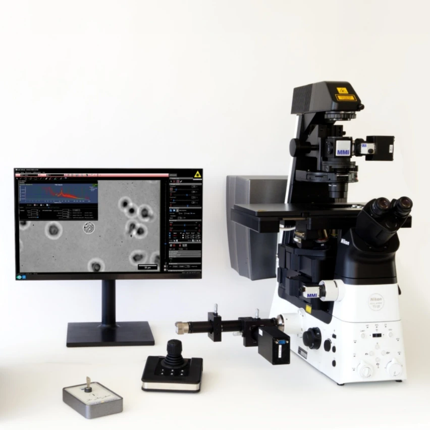
Mounted on Nikon Ti2
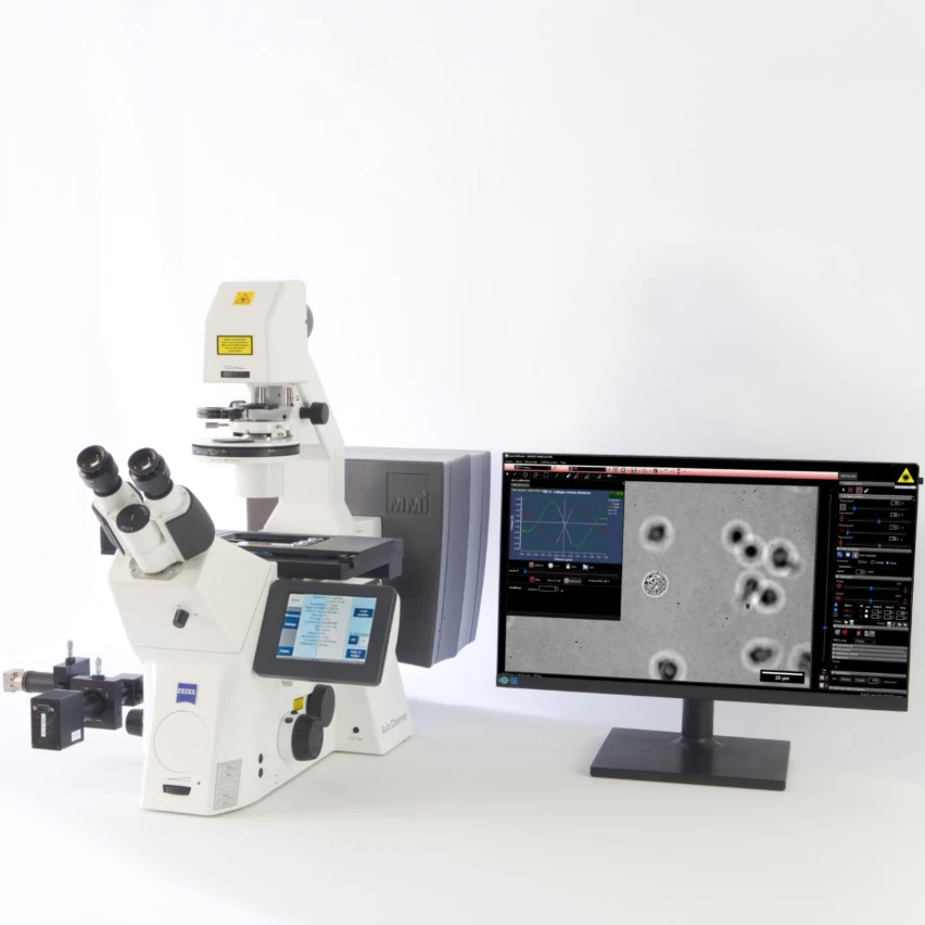
Mounted on Zeiss Axio Observer
Optical Tweezers are instruments that can hold and move microscopic particles just by employing laser light. Therefore, particles can be manipulated in a precise and contact-free way, only using the momentum generated by laser light. This makes optical tweezers a versatile and powerful tool for various research areas such as cell-based studies, measurement of binding forces as well as DNA or RNA binding.
Optical tweezers can optically trap microparticles that are transparent to infrared light and have an optical refraction index n of the object higher than that of the surrounding medium.
Yes, optical tweezers are ideally suited to study single cells. Using optical traps, the target cells can be manipulated in a precise and contact-free way, thereby avoiding any risks of contamination. In addition, using infrared lasers, the cell’s viability is not compromised during the experiment. Thus, the optical tweezer technology is also suitable for working with living cells, such as for drug discovery projects. Some optical tweezer systems, such as the MMI CellManipulator, are equipped with a second tweezer level to independently hold two cells or particles at the very same time and to precisely measure their interaction forces by force spectroscopy.
Find here a comprehensive overview of applications of optical tweezers in life science.