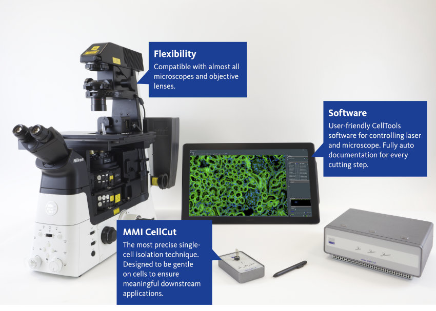
Laser Microdissection for highest sample integrity: MMI CellCut
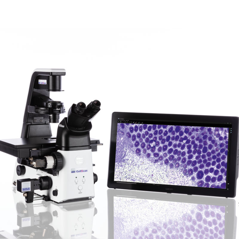
The MMI CellCut facilitates precise and contamination-free dissection of cell clusters, single cells or subcellular compartments from various types of tissues including fresh frozen, paraffin-embedded and archived slides, cytospins, smears and even living cells.

“I have compared all current available LMD platforms thoroughly and I got to say the CellCut plus is the most impressive one and I highly recommend it to our colleagues. Thanks for bring it to me.”
Chinese Academy of Sciences
Guangzhou, China
Have a look how it works
In the following video you will see how to select cells of interest, how to cut them and how you verify the successful collection
What is Laser Capture Microdissection?
The MMI CellCut provides a fully auditable and automatically documented workflow:

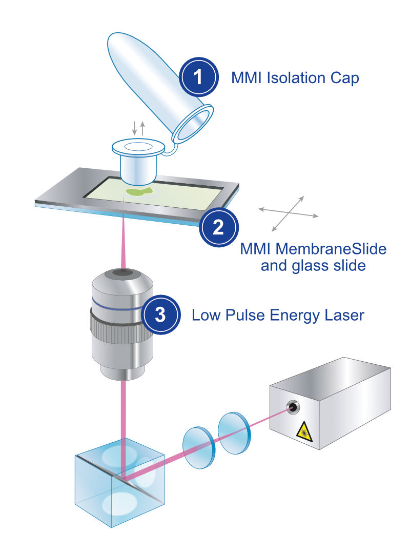
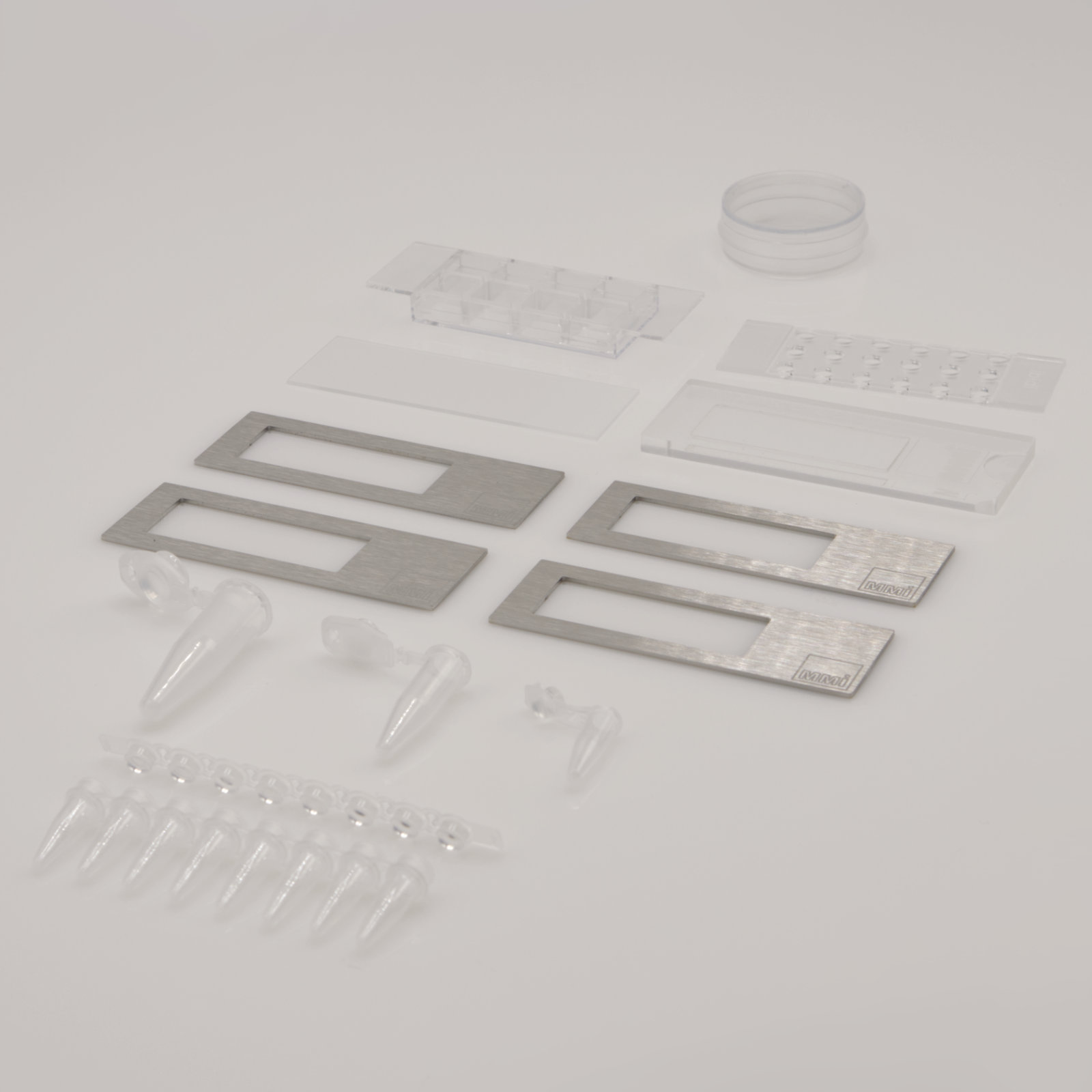
1. Sample Preparation: The tissue sample is first prepared for microdissection by fixing, embedding, and sectioning it into an inert membrane slide with negligible autofluorescence. Afterwards, the membrane slide is inverted and placed onto a glass slide for protection against contamination.
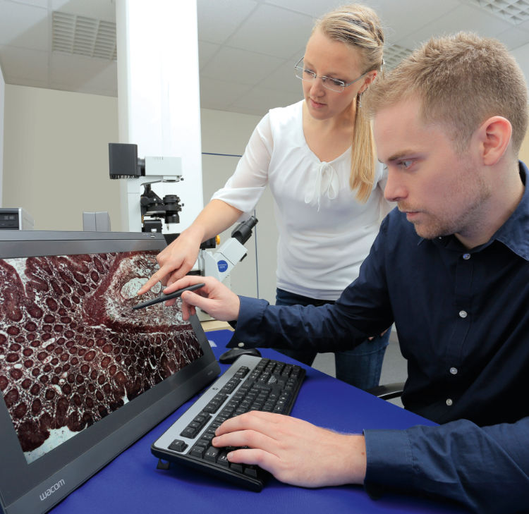
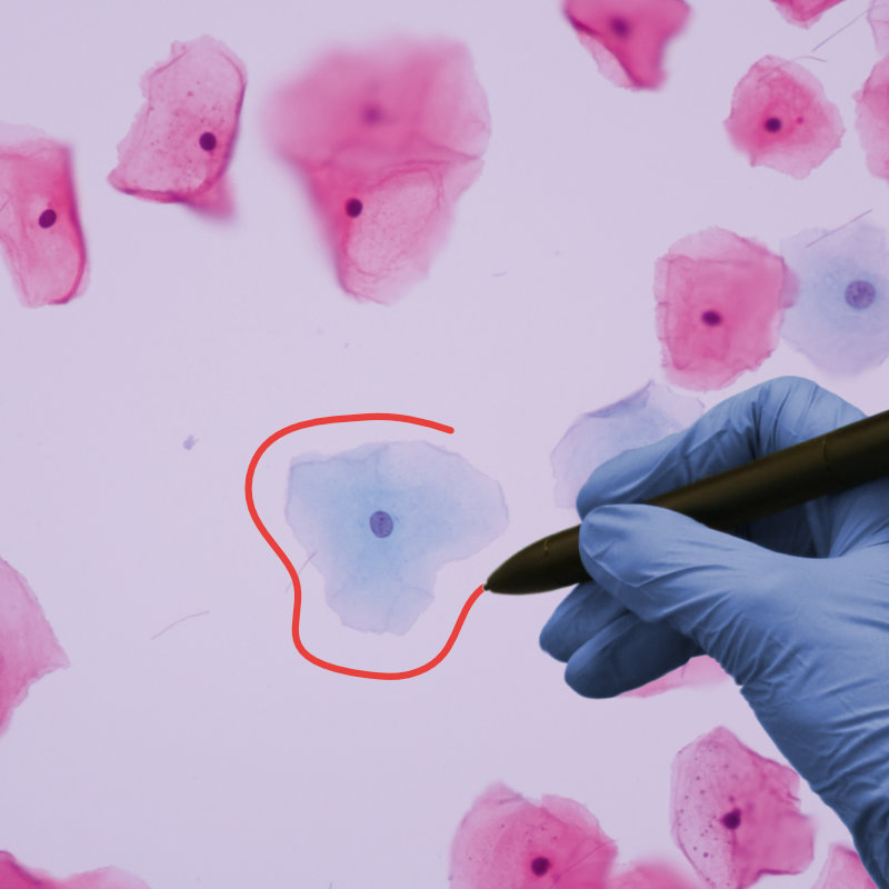
2. Cell Selection: The cells of interest can be selected on the computer screen using either the mouse, by freehand or by predefined geometrical shapes. Any number of cells across the slide can be identified as targets within one screening process.
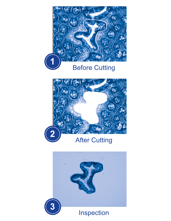
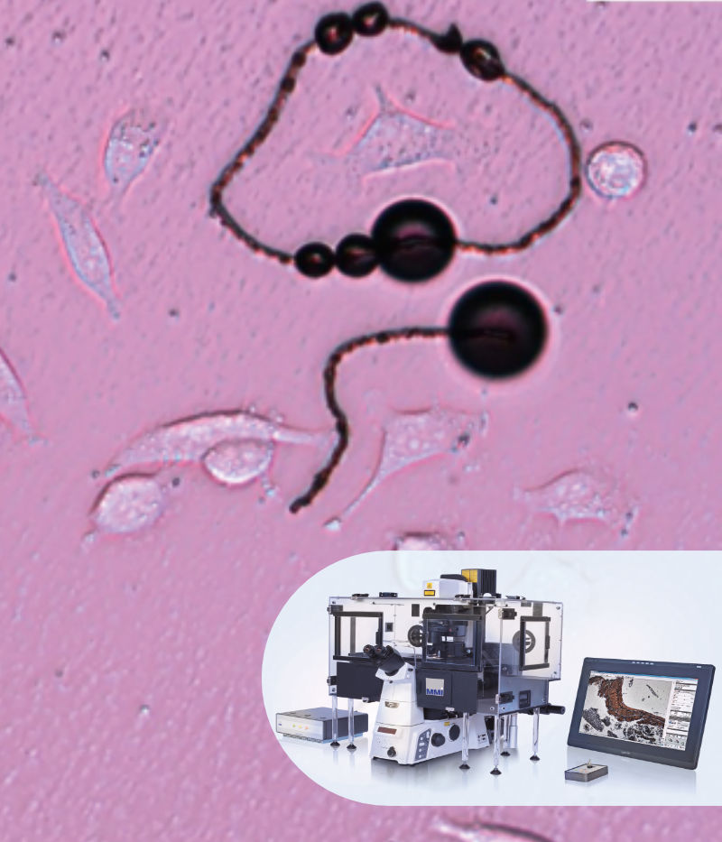
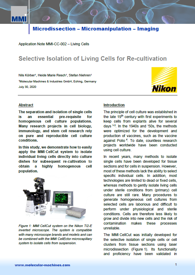
3. Laser Cutting: Once the region/cells of interest have been identified, the laser is focused on the selected area. The thin laser cutting path enables a precise and gentle extraction at an outstanding speed.
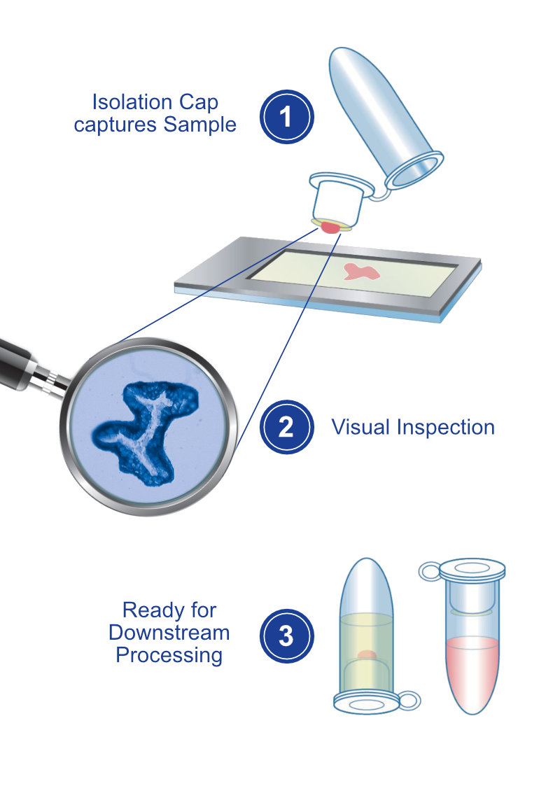
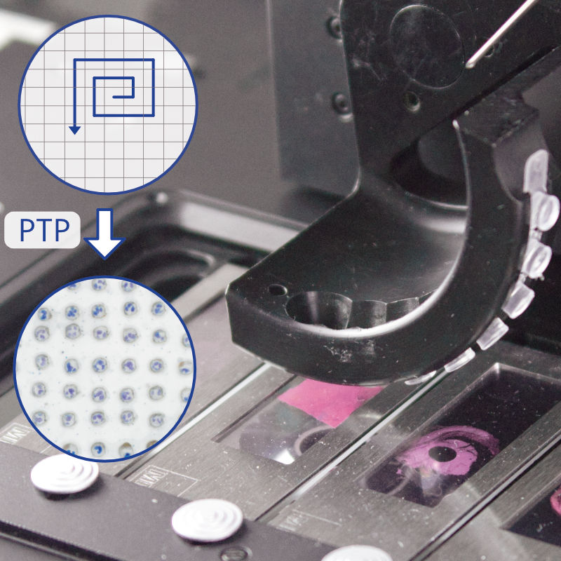
4. Collection & Inspection: The isolated target cell is collected by an adhesive cap. The orientation and morphology will be maintained. After cutting the cell can be visualised on the cap.
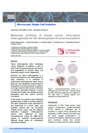
5. Ready for Downstream Processing: After microdissection, the collected cells can be processed further for any downstream application (e.g. gene expression analysis, RNA isolation and much more).
“We appreciate the resistant MMI product quality, the professional consulting, and the competent and quick service. MMI instruments are an important basis for our in-situ analysis in cellular tissue. The MMI CellCut laser microdissection followed by gene expression analysis is complementary for further routine methods like conventional optical microscopy (fluorescence), in-situ hybridization, and immunohistochemistry.”
RWTH Aachen
Germany

