

The most flexible and comprehensive whole slide imaging solution
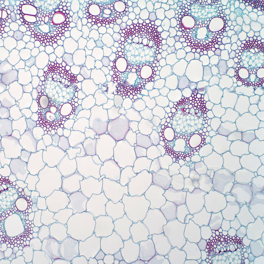
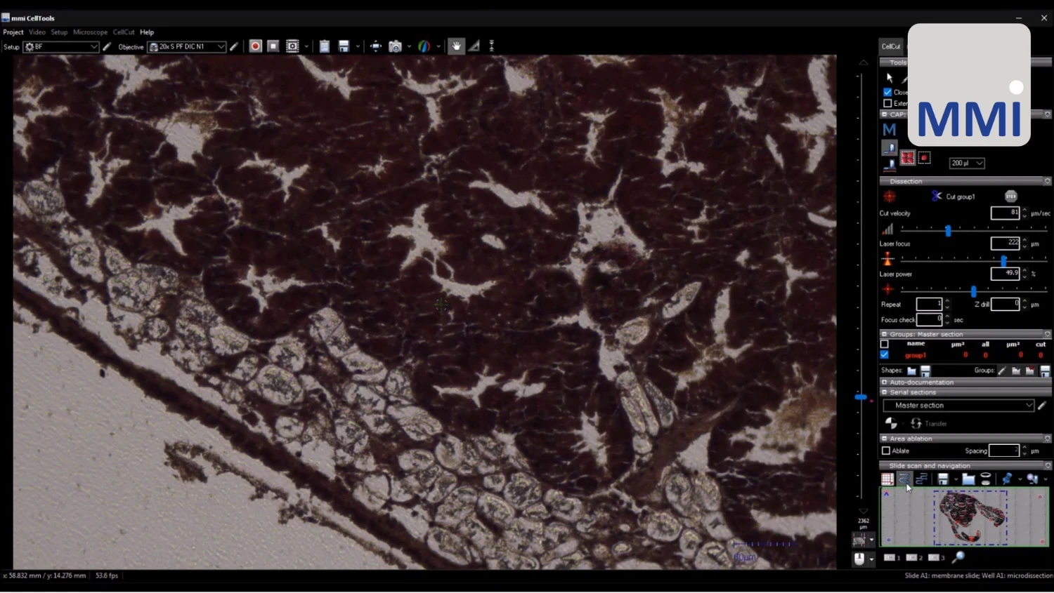
CellScan is compatible with all objectives, numerous research microscopes, and all possible imaging modes. Together with the MMI CellCut and CellDetector, it enables new areas of application, such as single cell isolation and automated cell analysis.
Whole Slide Scanning meets Laser Microdissection
By combining CellScan with CellCut, users can perform high-resolution whole slide imaging to accurately identify regions of interest (ROI) across the entire sample. Once identified, these specific target areas or single cells can be precisely isolated and collected using laser microdissection.
AI Cell Segmentation meets Whole Slide Scanning
Elevate your imaging workflows by pairing the CellScan with CellDetector. After acquiring the full slide scan, powerful AI-driven cell segmentation is applied to automatically identify specific cells or ROIs across the entire slide, drastically reducing manual effort and increasing analytical precision.
Watch our webinar to learn more: Webinar: Whole-Slide Imaging and Beyond
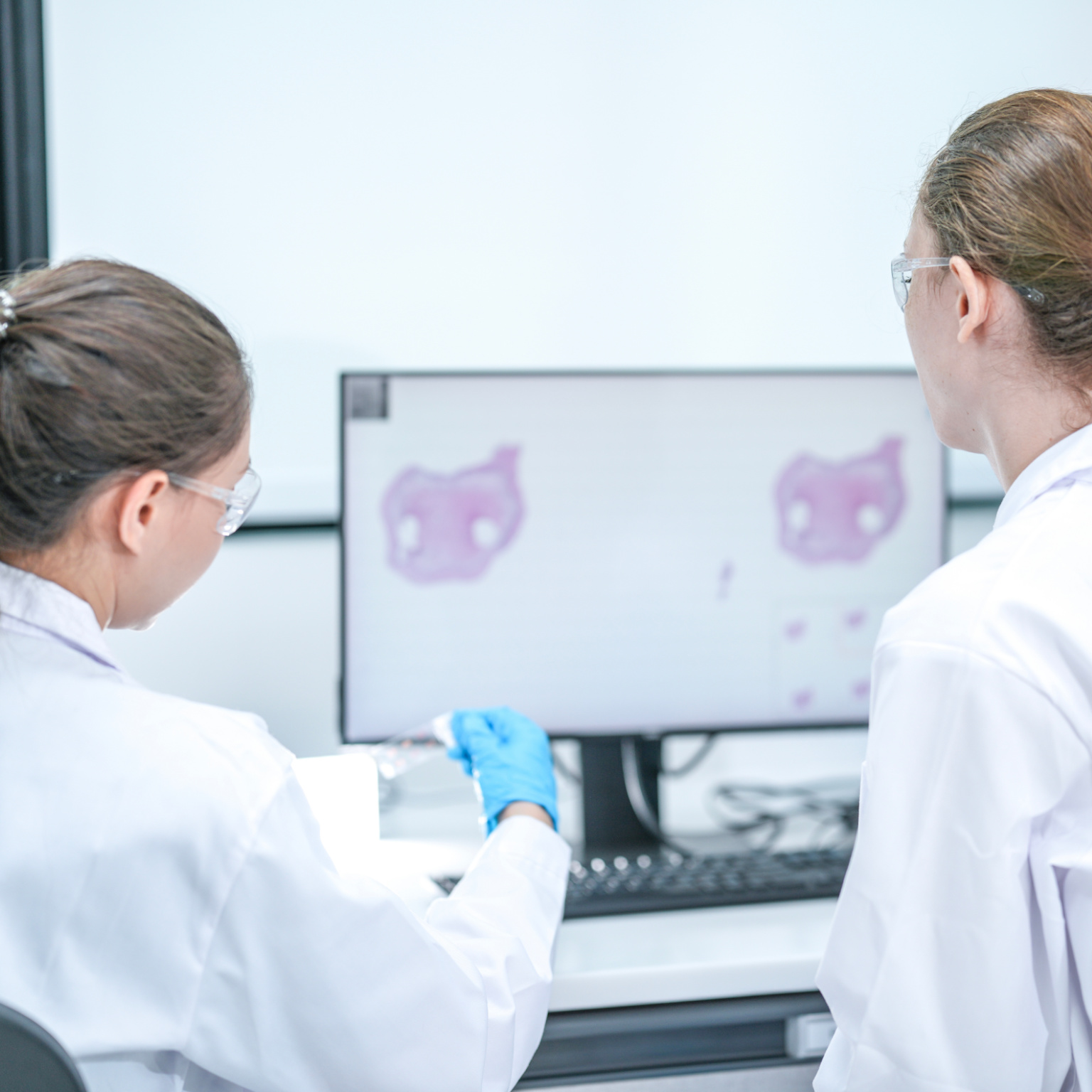
Unlock the potential of virtual microscopy and view your whole slide images remotely with unlimited licenses of the MMI imaging software CellViewer. Your digital images and slides can be stored, viewed, shared, and accessed globally. Collaborate with colleagues by sharing and virtually discussing your results.
Annotate, measure, and analyse seamlessly
High-quality imaging for a detailed view
Fast loading of BigTIFF files
Share whole-slide images with collaborators worldwide
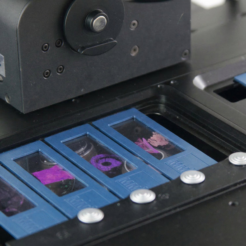
The entire imaging process is in your control, allowing you to scan a 15 mm x 15 mm section in less than 60 seconds and a full 75 mm x 25 mm slide in less than 7 minutes (20x objective).
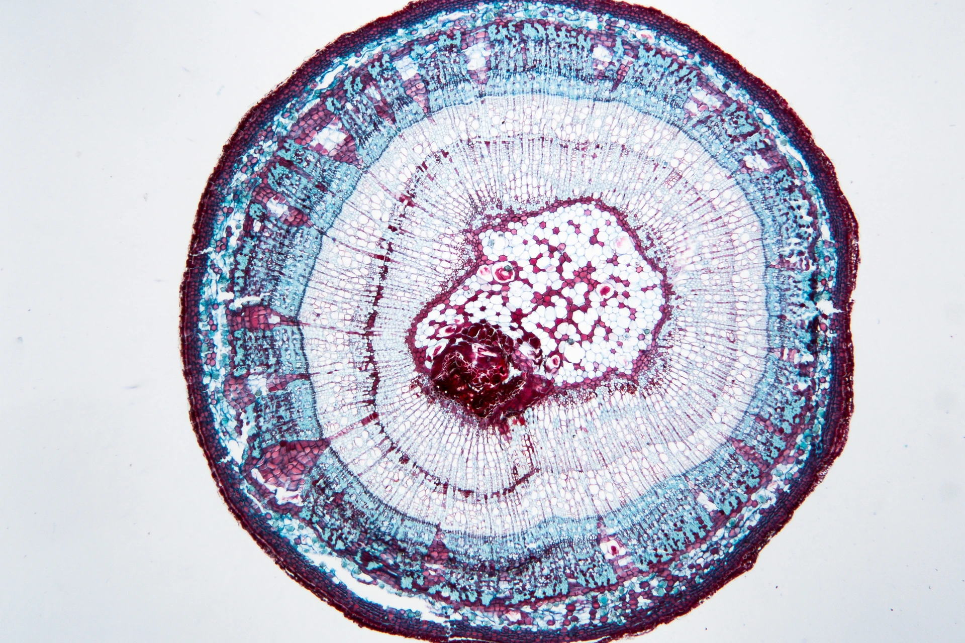
XY Imaging
Take full control of your research with CellScan’s powerful whole slide scanning capabilities. Seamlessly navigate across your entire samples using high-resolution, automated scanning along the X and Y axes, ensuring comprehensive and reliable coverage of every detail.
Z-Stack Imaging
Dive deeper into your specimens with advanced Z-stack imaging, enabling the precise exploration of complex structures across multiple focal planes. Instead of capturing just a single, flat image, our whole slide scanning system allows you to acquire a series of scans at consecutively adjusted Z-levels. This means you can digitally reconstruct the 3D architecture of thicker samples or tissue sections, ensuring that critical targets—regardless of their depth within the specimen—are always captured in perfect focus.
XYZ Scanning
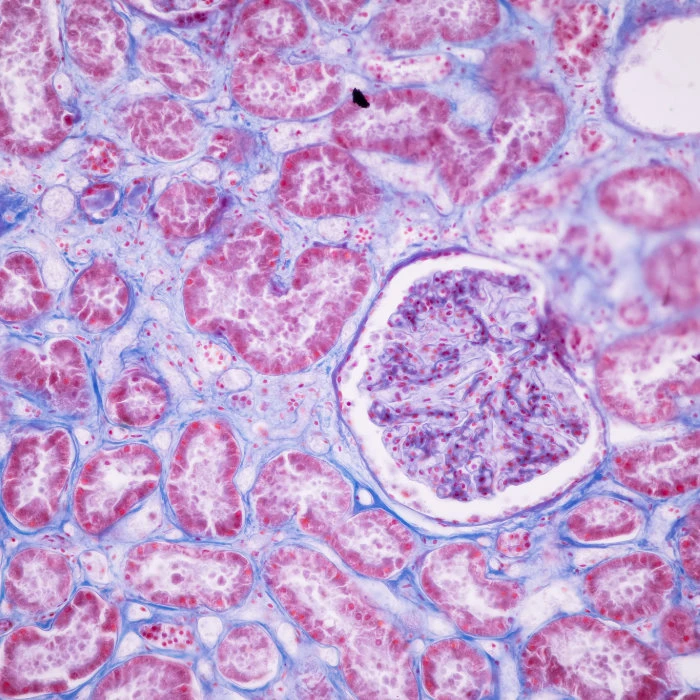
Comprehensive Multi-Fluorescence Imaging
Expand your analysis with multi-fluorescence imaging to visualise complex biological interactions with clarity and accuracy. Because the CellScan operates as a highly versatile whole slide scanner, it is fully capable of utilising all the advanced functions of your research microscope. Consequently, your scanning is never limited to a single resolution. Researchers can effortlessly change objectives or switch seamlessly between brightfield and fluorescence imaging modes with just a single mouse click.
Multi-Color Imaging
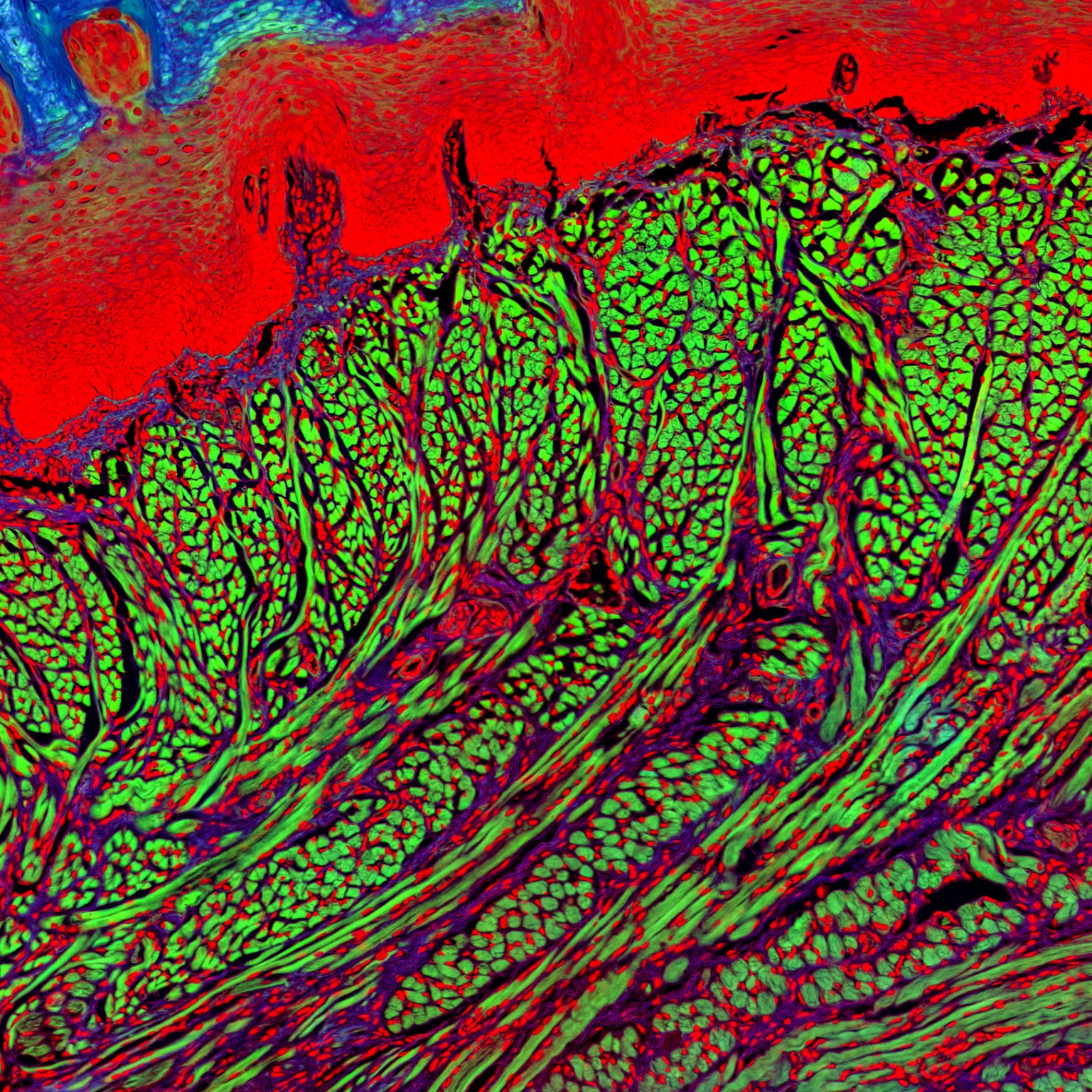
Live Cell Imaging
Live cell imaging involves time-lapse microscopy on living cells. Scientists use it to better understand biological function and cellular dynamics. With the whole slide scanner CellScan in combination with a stage-top incubator, you can easily perform live cell imaging experiments in fully physiological cell culture conditions.
Time-lapse
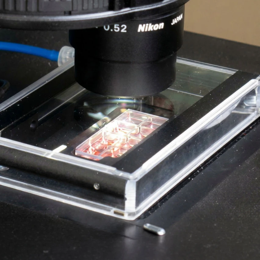
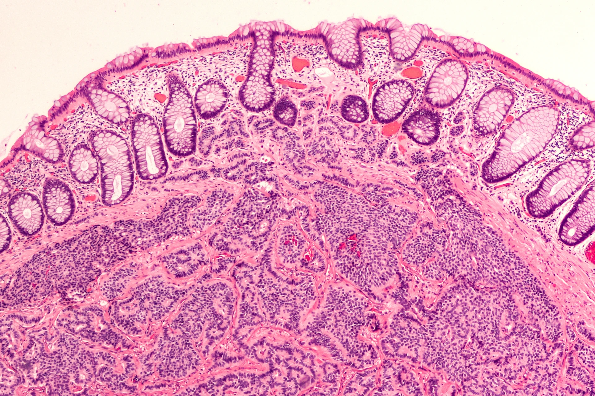
Thin skin showing the epidermis with its different strata, resting on the dermis
Whole Slide Imaging enables high-resolution scanning of entire tissue sections and forms the basis for efficient downstream analysis. In combination with the CellScan Whole Slide Scanner and the MMI CellCut laser microdissection system, it creates a streamlined workflow from imaging to precise sample isolation.
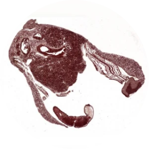
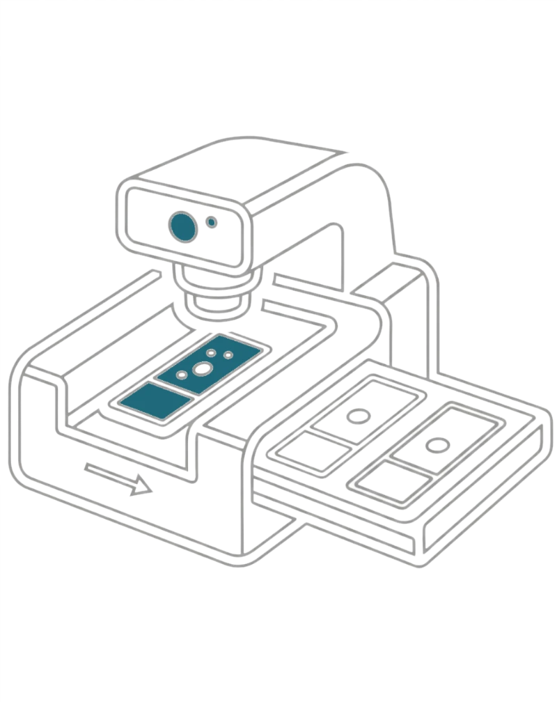
Scan your slides
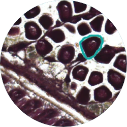
Identify the target region or use AI cell segmentation
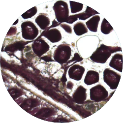
Isolate the target region
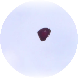
Verify successful isolation
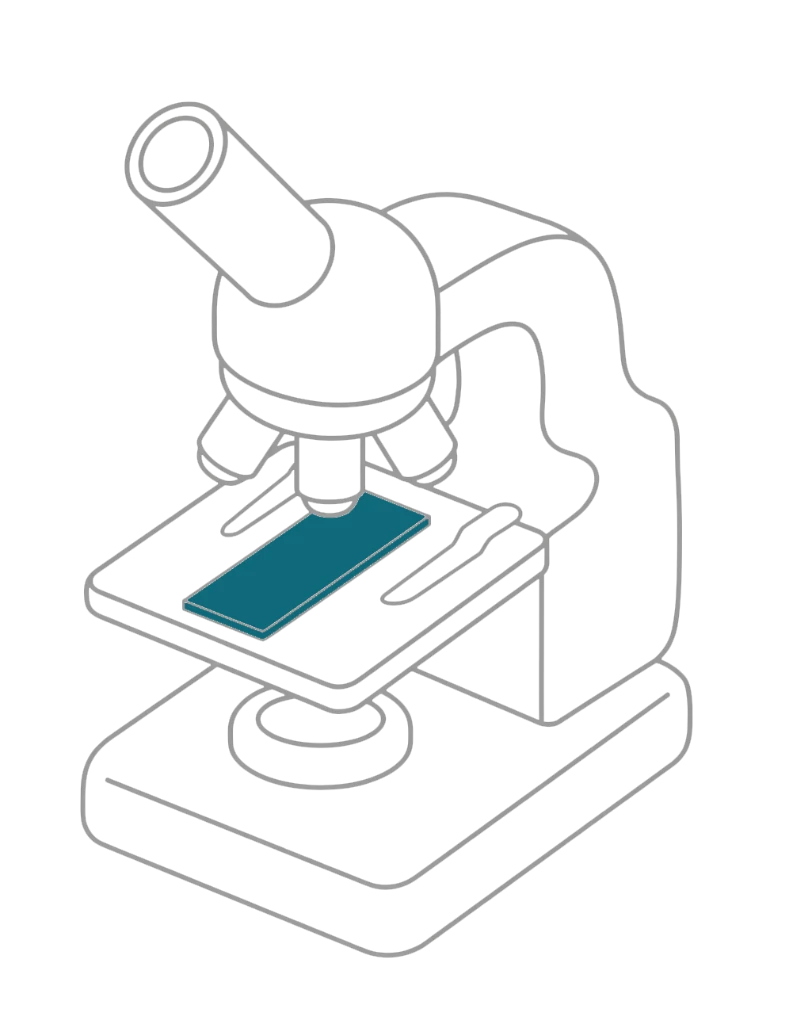
Anything you would do with a microscope
Any image mode, including confocal

With various microscope accessories
With any other MMI system (CellCut, CellEctor, CellDetector)
Watch how a slide is scanned with CellScan and how a single cell is isolated using the optional CellCut laser microdissection module.
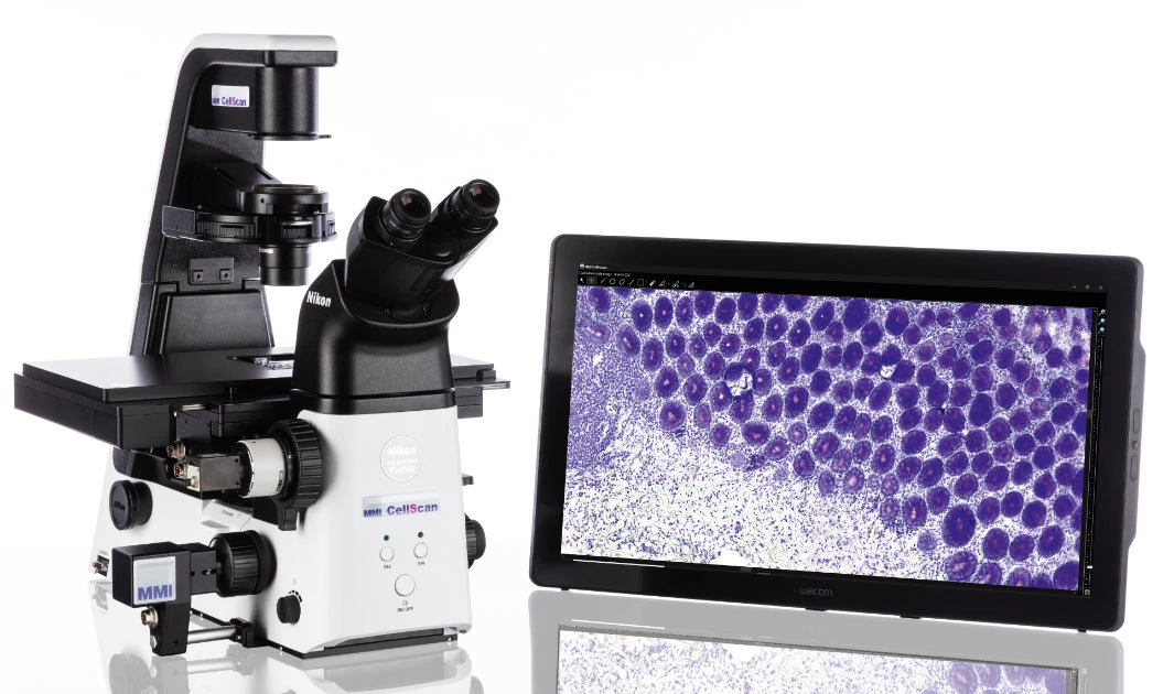
Compatible with almost all microscopes and imaging modes
The MMI CellScan is compatible with almost all research microscopes. Therefore, CellScan is not just a whole slide scanner, it’s a gateway to limitless digital imaging possibilities. Whether you’re working with brightfield, multi-fluorescence, confocal, phase contrast, or any other imaging mode, this versatile whole slide scanner gives you the freedom to explore and capture your samples in unprecedented detail.
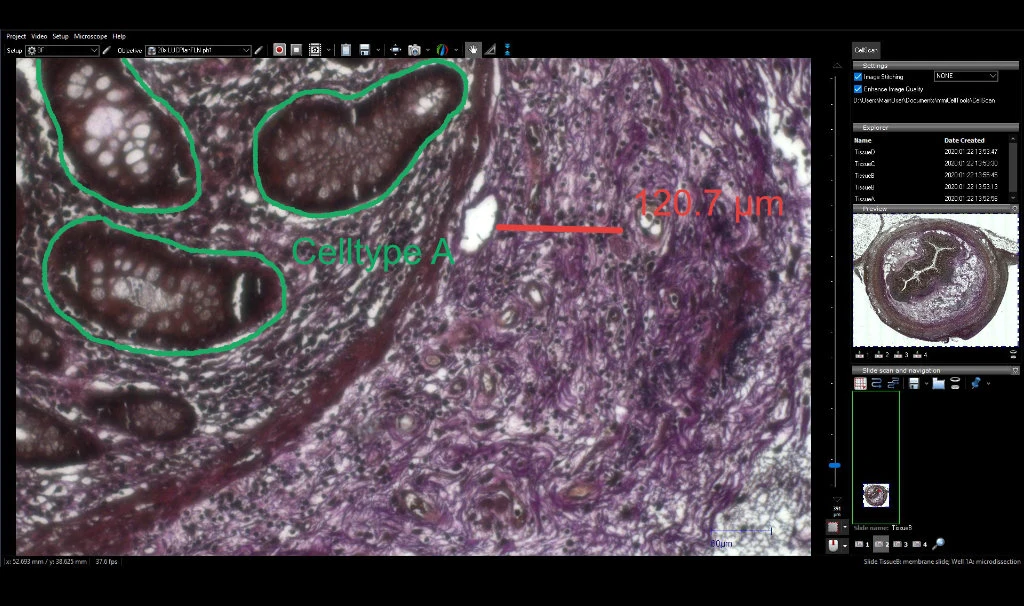
Capture and annotate
Mark and annotate cells and regions of interest effortlessly, with the added capability to export and reload marked areas. Insert scale bars, measure distances, and utilize this functionality for laser microdissection with the MMI CellCut. Capture videos and images at any time, providing flexibility in documentation.
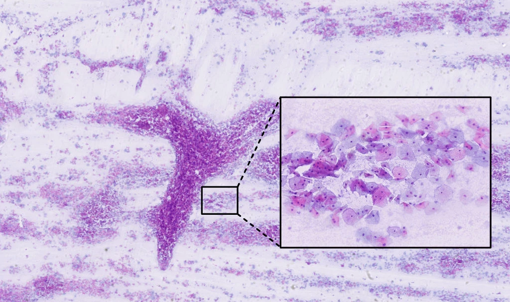
Stitching perfection for best image harmony
The MMI whole slide scanner boasts a sophisticated stitching function, ensuring a perfect alignment of individual images without any overlap or artefacts along the edges. This precision is achieved through the accuracy of the motorized stage, offering absolute positioning and sample localization. The stability of the microscope set-up, coupled with precise auto-focusing, particularly benefits samples with waviness along the z and focus directions.
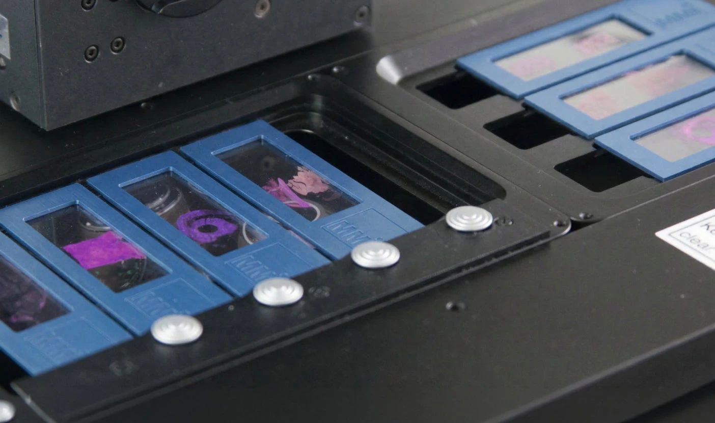
All common sample formats
The whole slide scanner CellScan supports various sample formats. From conventional glass slides (including coverslips) to petri dishes with living cells, and membrane slides designed for laser microdissection. This unmatched flexibility ensures that CellScan is tailored to meet the diverse needs of various applications, like in digital pathology, cell biology and oncology.
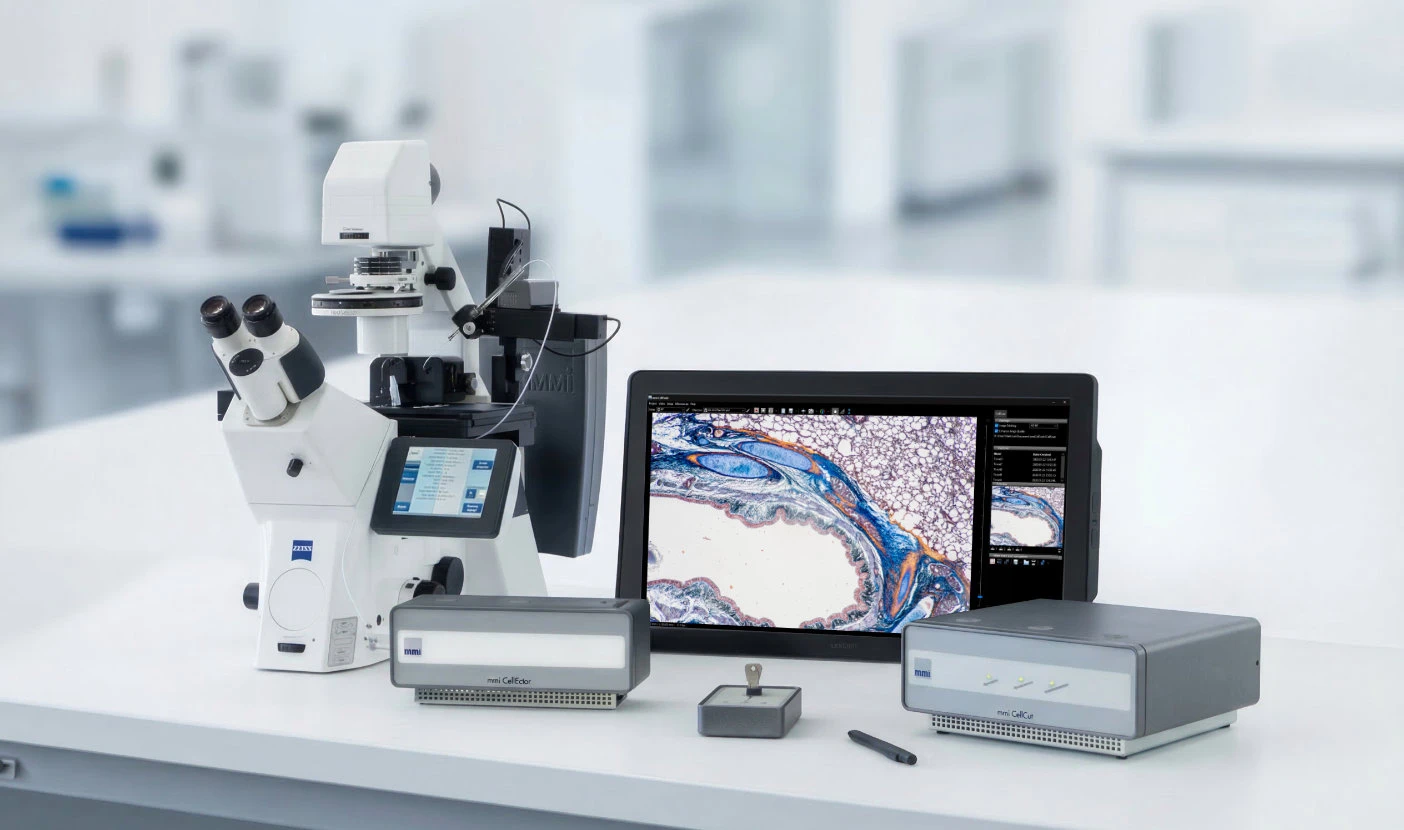
Modularity is key
The whole slide scanner CellScan boasts exceptional modularity, offering seamless compatibility and upgradability. With modules like CellCut for precise laser microdissection, CellEctor for isolating cells in suspensions, and CellDetector utilizing artificial intelligence for automatic cell and tissue identification, the whole slide scanner provides a versatile platform for evolving research needs.
Whole Slide Images are generated in BigTIFF format, enabling efficient storage and handling of very large image files.
A standard slide (75 mm × 25 mm) can be scanned in under 7 minutes using a 20x objective.
This depends on the stage being used. With the long travel stage, up to 8 slides can be scanned in sequence.
The CellScan whole slide scanner is compatible with almost all research microscopes.
Whole slide images can be edited using the free MMI CellViewer software. This software also allows users to import or draw shapes, which can be used for optional downstream laser microdissection with CellCut. Other software solutions can also be used, provided they support the BigTIFF format.
Whole slide imaging (WSI) – also known as virtual microscopy or whole slide scanning – is becoming increasingly popular in digital pathology as well as in clinical and basic research. A whole slide scanner can take images of each field of view across an entire microscopy slide. The pictures are then stitched together to generate a single, high-resolution digital whole slide image that researchers use for various purposes.
Whole slide imaging (WSI) offers significant advantages over conventional microscopy, including enhanced portability, seamless image sharing, efficient retrieval, improved workload distribution, and advanced image analysis capabilities.
Whole slide scanners are widely used across both clinical and nonclinical settings. In clinical practice, they are driving major advances in digital pathology, where studies have demonstrated a high level of agreement between diagnoses made using WSI and those based on traditional light microscopy.
In addition, biotechnology and pharmaceutical companies leverage whole-slide imaging to reduce costs, improve standardisation, and streamline data management throughout research and development processes.