
Spatial Omics
What is Spatial Omics?
Omics is a term summarizing various different analyses, such as genomics, epigenomics, transcriptomics, proteomics, lipidomics and metabolomics. Genomics, for example, analyzes the total genome, transcriptomics reads the total RNA and proteomics is evaluating the protein content. Spatial omics now combines the molecular analysis with spatial information on the cell’s localization within the tissue. Therefore, this technology is providing a powerful tool to understand important mechanisms such as tissue organization or cell regulation.
Single Cell Spatial Omics
The above mentioned methods, especially transcriptomics and proteomics, are currently being enhanced and optimized to achieve single cell resolution. Single cell spatial transcriptomics, for example, is a very powerful technology to understand the gene activity via RNA expression levels in each cell depending on its position and local environment within a tissue. Therefore, comparative transcriptomics compares different regions within a tissue regarding the transcriptional activity with detailed spatial resolution. In addition to single cell transcriptomics, the field of single cell proteomics is just emerging with new and improved technologies to detect proteins and peptides also in small sample volumes. Since not all RNA transcripts will be translated into stable proteins, this analysis provides further insights into a cell’s activity and status.
Publication News
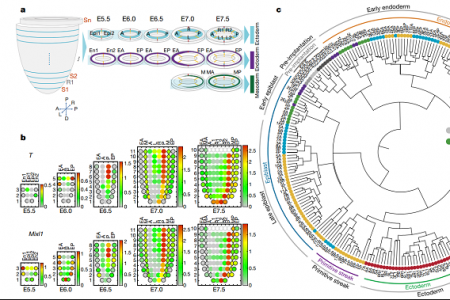
Read the latest publication by Dr. Guangdun Peng published in Nature.
How can you benefit from MMI technologies to perform single cell spatial omics analyses?
Laser microdissection is a technology employing microscopy to identify and select the cells of interest to be isolated. During isolation of distinct tissue areas or single cells by precise laser cutting, the tissue will stay intact and will not be dissociated into suspension cells. This method therefore maintains the spatial information of the samples to be analyzed and compared for their molecule content and activity. Thus, both information, the molecular information and the spatial information can be correlated to obtain a complete picture of the tissue.
Laser microdissection and spatial transcriptomics for example could visualize the gene expression level across a certain tissue.
MMI offers the MMI CellCut laser microdissection system for the precise cutting of tissue down to the single cell level.
Learn more about this technology here.
In 2018, MMI also developed the MMI CellScan for Whole Slide Imaging. This tool scans whole slides and saves them digitally in full resolution. Intriguingly, the MMI CellScan can be effectively combined with laser microdissection on a single microscopy platform. Therefore, images before and after cutting can be precisely correlated to downstream molecular analysis of the excised tissue areas or the excised single cells to allow for complete spatial omics analyses, such as spatial transcriptomics or spatial proteomics.
Learn more about the CellScan and its combination with the MMI CellCut.

“I have compared all current available LMD platforms thoroughly and I got to say the CellCut plus is the most impressive one and I highly recommend it to our colleagues. Thanks for bring it to me.”
Chinese Academy of Sciences
Guangzhou, China