

Optical Trapping
What can you do with optical trapping in Life Sciences?
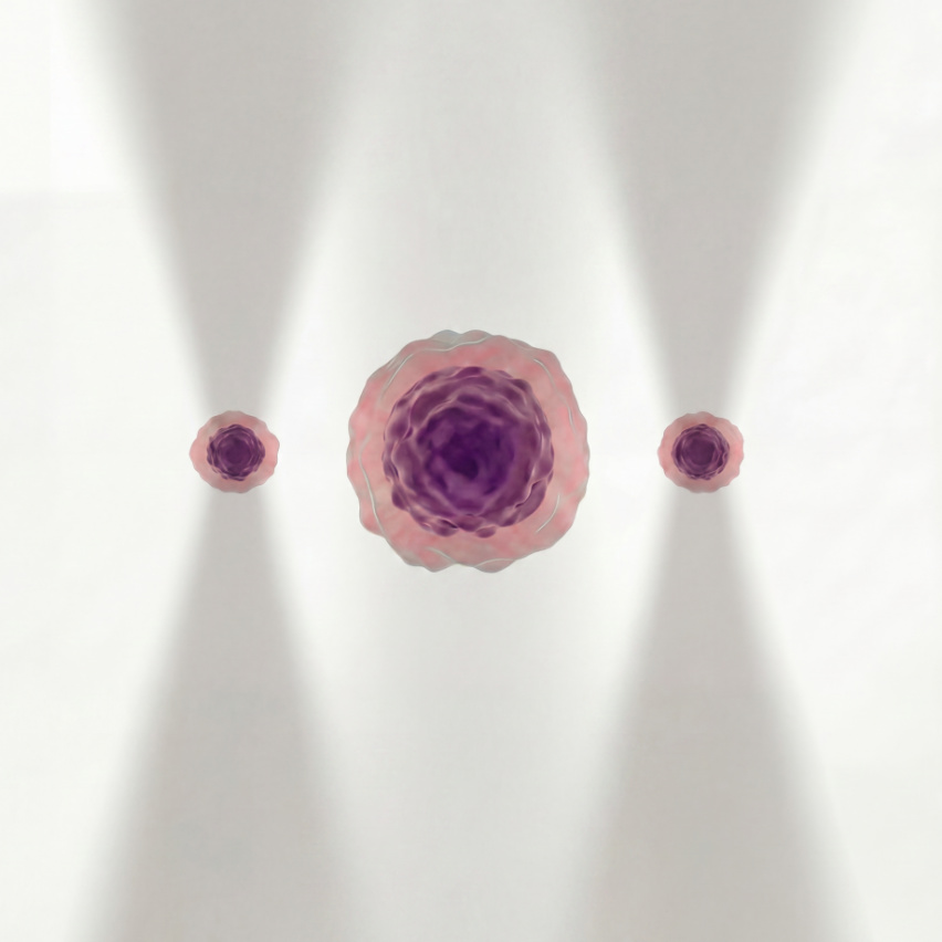
Motivation
Optical trapping is a very useful tool in life sciences that uses laser beams to grab and move tiny particles like beads, cells, organelles, DNA, and proteins. It helps scientific researchers to study how these particles behave and interact, allowing them to explore things at a much smaller level than before. Even though Optical Tweezers has many important uses in biology and medicine, not many people know about it or understand what it can do in life sciences.
This post is designed for readers who are either unfamiliar or somewhat familiar with the topic of optical trapping, providing a catalogue of optical trapping applications in life sciences. Every section contains several applications drawn from selected peer-reviewed publications.
Publications
Studying cell interactions is crucial for combating diseases because it helps us understand how cells communicate, cooperate, or conflict within the body. Many diseases, such as cancer, infections, and autoimmune disorders, result from disruptions in normal cell interactions. By examining how cells interact with each other and their environment, researchers can identify the underlying mechanisms that cause disease, develop targeted treatments, and improve drug delivery.
Here are several Optical Tweezers applications in studying cell interactions:
-
Immune Cell-Pathogen Interactions Study: Svoboda, K., Block, S. M. (1994). Biological applications of optical forces. Annual Review of Biophysics and Biomolecular Structure, 23, 247-285.
https://doi.org/10.1146/annurev.bb.23.060194.001335
-
Red Blood Cell Aggregation Study: Dao, M., Lim, C. T., & Suresh, S. (2003). Mechanics of the human red blood cell deformed by Optical Tweezers. Journal of the Mechanics and Physics of Solids, 51(11-12), 2259-2280.
https://doi.org/10.1016/j.jmps.2003.09.019
-
T-Cell and Antigen-Presenting Cell Interaction Study: Wei, X et al (1999). Mapping the sensitivity of T cells with an optical trap: Polarity and minimal number of receptors for Ca2+ signaling. PNAS, 96(15), 8471–8476.
1073/pnas.96.15.8471
-
Bacterial Adhesion to Host Cells Study: Ashkin, A., Dziedzic, J. M., & Yamane, T. (1987). Optical trapping and manipulation of single cells using infrared laser beams. Nature, 330(6150), 769-771.
1038/330769a0
-
Cell-Cell Adhesion in Cancer Metastasis Study: Xu, J., Tseng, Y., & Wirtz, D. (2000). Strain hardening of actin filament networks: Regulation by the dynamic cross-linking protein α-actinin. Journal of Biological Chemistry, 275(46), 35886-35892.
1074/jbc.M002377200
-
Phagocytic Cells – Microbe Interaction Study: Tam et al. (2010). Control and Manipulation of Pathogens with an Optical Trap for Live Cell Imaging of Intercellular Interactions. PLoS ONE 5(12): e15215.
https://doi.org/10.1371/journal.pone.0015215
-
Fibroblast – Fibronectin Coated Bead Interaction Study: Choquet et al (1997). Extracellular Matrix Rigidity Causes Strengthening of Integrin–Cytoskeleton Linkages. Cell. Vol 88, Issue 1, P39-48.
https://doi.org/10.1016/S0092-8674(00)81856-5
-
Sperm – Egg Interaction Study: Clement-Sengewald et al (1996). Fertilization of Bovine Oocytes Induced Solely with Combined Laser Microbeam and Optical Tweezers. Journal of Assisted Reproduction and Genetics, Vol. 13, No. 3, 259-265. https://doi.org/10.1007/BF02065947
-
Human Natural Killer Cells (NK) – Human Erytholeukemia (K562) Interaction Study: Seeger et al (1991). Application of laser Optical Tweezers in immunology and molecular genetics. Cytometry, 12(6):497-504.
1002/cyto.990120606
-
Cilia – Fluid Flow Interaction (Cell – Environment Interaction) Study: Katoh et al (2023). Immotile cilia mechanically sense the direction of fluid flow for left-right determination. Science, Vol. 379, Issue 6627 page 66-71.
DOI: 10.1126/science.abq8148
The ability to manipulate cell organelles is crucial to understand disease mechanisms. Diseases like neurodegenerative disorders and cancers are linked to organelle dysfunction. Researchers can use optical trapping to model dysfunctions by altering organelle position, providing insights into how diseases develop at the cellular level.
-
Plant Organelles Manipulation Study: Hawes et al (2010). Optical Tweezers for the micromanipulation of plant cytoplasm and organelles. Curr Opin Plant Biol. 13(6):731-5.
10.1016/j.pbi.2010.10.004
-
Golgi Bodies Manipulation in Plant Cells Study: Sparkes et al (2009). Grab a Golgi: laser trapping of Golgi bodies reveals in vivo interactions with the endoplasmic reticulum. Traffic. 10(5):567-71.
10.1111/j.1600-0854.2009.00891.x
-
Chloroplast Manipulation in Plant Cells Study: Bayoudh et al (2001). Micromanipulation of chloroplasts using Optical Tweezers. J Microsc. 203(Pt 2):214-22.
10.1046/j.1365-2818.2001.00843.x
-
Amyloplast Manipulation in Plant Cells Study: Abe et al (2020). Micromanipulation of amyloplasts with Optical Tweezers in Arabidopsis stems. Plant Biotechnol (Tokyo). 37(4): 405-415.
10.5511/plantbiotechnology.20.1201a
-
Peroxisome Manipulation in Plant Cells Study: Gao et al (2016). In Vivo Quantification of Peroxisome Tethering to Chloroplasts in Tobacco Epidermal Cells Using Optical Tweezers. Plant Physiology. Vol. 170, Issue 1, Pages 263-272. https://doi.org/10.1104/pp.15.01529
-
Neuronal Mitochondria Manipulation Study: Gong et al (2023). Optical manipulation of neuronal mitochondria using scanning Optical tweezers. Journal of Innovative Optical Health Science, 98(2).
https://dx.doi.org/10.1142/S1793545823410018
-
Transfer Mitochondria from Fetal to Adult Mesenchymal Stem Cells Study: Shakoor et al (2021). Automated Optical Tweezers Manipulation to Transfer Mitochondria from Fetal to Adult MSCs to Improve Antiaging Gene Expressions. Small.
-
Controlling Nuclei Positioning Study: Du et al (1991). Laser tweezer manipulation of micronuclei in Paramecium. OSA Annual Meeting 1991.
https://doi.org/10.1364/OAM.1991.WG5
-
Chromosomes and Mitotic Spindle Manipulation Study: Berns et al (2006). Chapter 36 – Micromanipulation of Chromosomes and the Mitotic Spindle Using Laser Microsurgery (Laser Scissors) and Laser-Induced Optical Forces (Laser Tweezers). Cell Biology (3rd Ed.) A Laboratory Handbook. Vol.3, pages 351-363.
https://doi.org/10.1016/B978-012164730-8/50164-7
-
Manipulation of Filamentous Fungi Organelles Study: Wright et al (2007). Optical tweezer micromanipulation of filamentous fungi. Fungal Genetics and Biology. 44, pages 1-13.
https://doi.org/10.1016/j.fgb.2006.07.002
Using optical trapping in vivo offers a non-invasive, precise, and highly controlled way to study complex biological systems. It opens new possibilities for investigating the physical forces and interactions driving essential life processes, all while maintaining the integrity of the living organism.
-
Cell Manipulation Inside Living Zebrafish Study: Johansen et al. (2016). Optical micromanipulation of nanoparticles and cells inside living zebrafish. comm. 7:10974.
DOI: 10.1038/ncomms10974
-
Manipulating Injected Fluorescent Beads Inside Living Zebrafish Embryos Study: Hoerner et al. (2017). Holographic Optical Tweezers-based in vivo manipulations in zebrafish embryos. Journal of Biophotonics.
https://doi.org/10.1002/jbio.201600226
-
Trap RBC in Living Mice Study: Zhong et al. (2013). Trapping red blood cells in living animals using Optical Tweezers. Nature Communications. 1768 (2013).
https://doi.org/10.1038/ncomms2786
-
Measuring Flow and Drag Forces Imposed to Trapped RBC of Living Zebrafish Embryos Study: Harleep et al. (2017). Hemodynamic forces can be accurately measured in vivo with Optical Tweezers. Mol Bio Cell. 28(23): 3252–3260.
10.1091/mbc.E17-06-0382
-
Probing Cell Mechanics in Drosophila Embryo Study: Chardes et al. (2017). Probing Cell Mechanics with Bead-Free Optical Tweezers in the Drosophila Embryo. J Vis Exp. 2:141.
10.3791/57900
Studying giant unilamellar vesicles (GUVs) with optical trapping offers valuable insights into membrane biophysics, cell mechanics, and molecular interactions. GUVs are artificial, cell-sized vesicles composed of a single lipid bilayer, which makes them excellent model systems for investigating the properties and behaviour of biological membranes in a controlled environment. Optical trapping provides precise, non-invasive manipulation of GUVs, making them an ideal tool for studying these vesicles.
-
Generating Membrane Nanotubes from Lipid Vesicles Study: Prévost et al. (2017). Pulling Membrane Nanotubes from Giant Unilamellar Vesicles. J. Vis. Exp. (130), e56086, doi:10.3791/56086.
10.3791/56086
-
Generating Long Tubular Nanostructures Resembling Biological Membranes from GUV Study: Roux et al. (2005). Role of curvature and phase transition in lipid sorting and fission of membrane tubules. The EMBO Journal. 24(8):1537-1545.
10.1038/sj.emboj.7600631
-
Characterization of membrane protein dynamics using GUV and SUV Study: Wang et al. (2021). Single-molecule manipulation of macromolecules on GUV or SUV membranes using Optical Tweezers. Biophys J. 120(24): 5454-5465,
10.1016/j.bpj.2021.11.2884
-
Stretching GUV Study: Solmaz et al. (2021). Optical stretching of giant unilamellar vesicles with an integrated dual-beam optical trap. Biomed Opt Express. 3(10): 2419-2427,
10.1364/BOE.3.002419
-
Sculpting and fusing biomimetic vesicle networks Study: Bolognesi et al. (2018). Sculpting and fusing biomimetic vesicle networks using Optical Tweezers. Nature Communications. 1882,
https://doi.org/10.1038/s41467-018-04282-w
Optical trapping is particularly useful for measuring cellular binding or interaction forces because they allow researchers to quantify the weak, dynamic forces involved in molecular and cellular interactions with high precision. By trapping and manipulating individual cells or biomolecules, Optical Tweezers can measure the forces required to form or break molecular bonds, such as those involved in receptor-ligand interactions, cell adhesion, or immune responses. This technique enables real-time observation of binding events and interaction dynamics, providing critical insights into cellular processes. Importantly, because optical trapping is non-invasive, it preserves cell viability and function, allowing for the study of living cells under near-physiological conditions.
-
Human Bone Cells and Implant Surfaces Study: Andersson et al. (2007). Using Optical Tweezers for measuring the interaction forces between human bone cells and implant surfaces: System design and force calibration. Sci. Instrum. 78, 074302. https://doi.org/10.1063/1.2752606.
-
Motility Forces of Isolated Chromosomes Study: Khatibzadeh et al (2014). Determination of motility forces on isolated chromosomes with laser tweezers. Sci Rep, 4:6866.
10.1038/srep06866
-
Ligand – Receptor Binding in Living Cells Study: Riesenberg et al (2020). Probing Ligand-Receptor Interaction in Living Cells Using Force Measurements with Optical Tweezers. Front. Bioeng. Biotechnol. Vol.8.
https://doi.org/10.3389/fbioe.2020.598459
-
Fibrinogen – Integrin Binding in Living Cells Study: Litvinov et al (2002). Binding strength and activation state of single fibrinogen-integrin pairs on living cells. PNAS. 99(11)7426-7431.
https://doi.org/10.1073/pnas.11219499
-
Determining position-dependent linkages of multiple proteins. Study: Nishizaka et al (2000). Position-dependent linkages of fibronectin– integrin–cytoskeleton. PNAS. 97(2)692-697.
https://doi.org/10.1073/pnas.97.2.692
Optical trapping is highly useful for mechanical characterisation because they enable precise manipulation and measurement of microscopic particles without physical contact. By using a focused laser beam to trap and move small objects such as cells, biomolecules, or nanoparticles, optical trapping can exert finely controlled forces in the pico- to nanonewton range. This allows researchers to study the mechanical properties of materials, like stiffness, elasticity, or molecular interactions, with exceptional precision. Additionally, their non-invasive nature ensures minimal damage to delicate samples, making them ideal for biological systems and soft matter, where conventional tools may be too disruptive.
-
Leukemia Cells Study: Zhou et al (2014). Accurate Measurement of Stiffness and Leukemia Cells and Leukocytes Using an Optical Trap by a Rate-Jump Method. RSC Advance, 4, 8453-8460.
https://doi.org/10.1039/C3RA45835K
-
Cytoplasm Stiffness in Plant Cells Study: van der Honing et al (2010). Actin and myosin regulate cytoplasm stiffness in plant cells: a study using Optical Tweezers. New Phytol. 185(1):90-102.
1111/j.1469-8137.2009.03017.x
-
Drug-treated Jurkat Cells Study: Khakshour et al (2010). Mechanical characterization of ART-treated Jurkat cells using Optical Tweezers. Annu Int Conf IEEE Eng Med Biol Soc. 6806-9.
1109/EMBC.2014.6945191
-
Cancer Cells under Hypoxia Study: Khakshour et al (2017). Retinoblastoma protein (Rb) links hypoxia to altered mechanical properties in cancer cells as measured by an Optical Tweezers. Sci Rep. 7(1):7833.
1038/s41598-017-07947-6
-
Red Blood Cells Elasticity in response to Antimalarials Study: Dorta et al (2017). Optical Tweezers to measure the elasticity of red blood cells: a tool to study the erythrocyte response to antimalarials. Malar. Vol 2.
https://doi.org/10.3389/fmala.2024.1362644
-
The Stiffness of Alginate-Gelatin Hydrogel Study: Ling et al (2022). A Novel Step-T-Junction Microchannel for the Cell Encapsulation in Monodisperse Alginate-Gelatin Microspheres of Varying Mechanical Properties at High Throughput. 12(8):659.
10.3390/bios12080659
-
Membrane Cell Tension Study: Kreysing et al (2022). Effective cell membrane tension is independent of polyacrylamide substrate stiffness. PNAS Nexus. 2(1):pgac299.
1093/pnasnexus/pgac299
-
Rheological Properties of Fibroblasts, Neurons, and Astrocytes Study: Ayala et al (2016). Rheological properties of cells measured by Optical Tweezers. BMC Biophysics. 9:5.
https://doi.org/10.1186/s13628-016-0031-4
-
Viscosity of out-of-equilibrium System Study: Madsen et al (2021). Ultrafast viscosity measurement with ballistic Optical Tweezers. Nature Photonics. 15, 386-392.
https://doi.org/10.1038/s41566-021-00798-8
-
Ex Vivo Generated Red Blood Cells Study: Bernecker et al (2021). Biomechanics of Ex Vivo-Generated Red Blood Cells Investigated by Optical Tweezers and Digital Holographic Microscopy. 10(3):552.
10.3390/cells10030552
Molecular motors, such as myosin, kinesin, and dynein, are proteins that convert chemical energy into mechanical work, driving processes like muscle contraction, intracellular transport, and cell division. To understand how these motors function at the molecular level, researchers need a tool that can exert controlled forces on microscopic particles while observing their movement in real-time — this is where optical trapping excels.
-
Microtubule Translocation by Single Kinesin Study: Kuo and Sheetz (1993). Force of single kinesin molecules measured with Optical Tweezers. 260(5105):232-4. 10.1126/science.8469975
-
Kinetochore – Microtubule Interaction Study: Muir et al (2023). Structural mechanism of outer kinetochore Dam1-Ndc80 complex assembly on microtubules. Vol 382, No. 6675 pp 1184-1190.
DOI: 10.1126/science.adj8736
-
Single Kinesin Moving a Bead Study: Block et al (2007). Bead movement by single kinesin molecules studied with Optical Tweezers. Nature. 348(6299):348-52.
1038/348348a0
-
F-Actin and Titin Interaction Study: Bianco et al (2007). Interaction Forces between F-Actin and Titin PEVK Domain with Optical Tweezers. Biophysical Journal. 93, Issue 6, P2102-2109.
https://doi.org/10.1529/biophysj.107.106153
-
Single Myosin Molecule – Single Suspended F-actin Interaction Study: Finer et al (2007). Single Myosin Molecule Mechanics: Piconewton Forces and Nanometre Steps. Nature. 368, 113-119.
https://doi.org/10.1038/368113a0
-
Myosin V stepping on F-actin Study: Capello et al (2007). Myosin V Stepping Mechanism. PNAs. 104(39)15328 – 15333. https://doi.org/10.1073/pnas.0706653104
-
Determining the Working Stroke of Rabbit Heavy Meromyosin Study: Veigl et al (2007). The Stiffness of Rabbit Skeletal Actomyosin Cross-Bridges Determined with an Optical Tweezers Transducer. Journ. Vol. 75, Issue 3, 1424-1438.
https://doi.org/10.1016/S0006-3495(98)74061-5
-
Dynein – Microtubule Interaction Study: Brenner et al (2007). Force production of human cytoplasmic dynein is limited by its processivity. Adv. Vol. 6, No 15.
DOI: 10.1126/sciadv.aaz4295
-
Talin – Integrin Bond Study: Bodescu et al (2007). Kindlin stabilizes the talin·integrin bond under mechanical load by generating an ideal bond. 120(26):e2218116120.
10.1073/pnas.2218116120
-
Single Fibrinogen – Integrin Pair Bond Study: Litvinov et al (2002). Binding strength and activation state of single fibrinogen-integrin pairs on living cells. 99 (11) 7426-7431.
https://doi.org/10.1073/pnas.112194999
Optical trapping is highly valuable for studying DNA or RNA interaction forces because they enable precise manipulation of these delicate molecules to measure the forces involved in processes such as folding, unfolding, and binding with proteins or other nucleic acids. By trapping and stretching individual DNA or RNA strands, researchers can directly observe mechanical responses to applied forces, providing insights into their structural stability and interactions with enzymes like polymerases or helicases. This technique allows for real-time, single-molecule measurements of molecular interactions, which is critical for understanding the mechanics of transcription, replication, and gene regulation. The ability to perform these studies without damaging the nucleic acids ensures accurate, biologically relevant results.
-
Unzipping DNA Study: Bockelmann et al (2002). Unzipping DNA with Optical Tweezers: High Sequence Sensitivity and Force Flips. Journ. Vol. 82, Issue 3, P1537-1553.
10.1016/S0006-3495(02)75506-9
-
Stretching DNA Study: Wang et al (1997). Strecthing DNA with Optical Tweezers. Biophys Mar; 72(3): 1335–1346. https://doi.org/10.1016%2FS0006-3495(97)78780-0
-
Coiled coil force sensor coupled to DNA handle Study: Ren et al (2023). Force redistribution in clathrin-mediated endocytosis revealed by coiled-coil force sensor. Sci Adv. 9(41):eadi1535.
1126/sciadv.adi1535
-
Using DNA as Standard for Length and Force Measurements Study: Rickgauer et al (2006). DNA as a Metrology Standard for Length and Force Measurements with Optical Tweezers. Vol. 91, Issue 11, 4253-4257.
https://doi.org/10.1529/biophysj.106.089524
-
DNA-protein-DNA Force Extension, Manipulation, and Visualization Study: Brouwer et al (2017). Human RAD52 Captures and Holds DNA Strands, Increases DNA Flexibility, and Prevents Melting of Duplex DNA: Implications for DNA Recombination. Vol. 18, Issue 12, 2845-2853.
https://doi.org/10.1016/j.celrep.2017.02.068
-
Observing Mechano-Chemical Cycles of DNA Packaging by φ29 Motor with Base Pair Resolution Study: Chistol et al (2017). High Degree of Coordination and Division of Labor among Subunits in a Homomeric Ring ATPase. Vol. 151, Issue 5, P1017-1028. https://doi.org/10.1016/j.cell.2012.10.031
-
Observing How Biomolecular Condensation Physically Prevents DNA End Disjunction Study: Chappidi et al (2024). PARP1-DNA co-condensation drives DNA repair site assembly to prevent disjunction of broken DNA ends. Vol. 187, 945-961. https://doi.org/10.1016/j.cell.2024.01.015
-
Studying RNA-Protein Interactions in Translation Regulation Study: Pekarek et al (2022). Optical Tweezers to Study RNA-Protein Interactions in Translation Regulation. J Vis Exp. 12:180.
https://doi.org/10.3791/62589
-
Observing RNA Force Unfolding Kinetics Study: Wen et al (2007). Force Unfolding Kinetics of RNA Using Optical Tweezers. I. Effects of Experimental Variables on Measured Results. Journ. Vol. 92, Issue 9 P2996-3009.
https://doi.org/10.1529/biophysj.106.094052
-
Studying the Transcription by Eukaryotic RNA Polymerases Study: Lisica and Grill (2017). Optical Tweezers Studies of Transcription by Eukaryotic RNA Polymerases. Biomolecular Concepts. Vol. 8, Issue 1.
https://doi.org/10.1515/bmc-2016-0028
Combining Optical Tweezers with Raman spectroscopy is highly useful because it enables simultaneous manipulation and molecular analysis of small particles or biological molecules with high precision.
-
Comparing the Deformability of Human vs Camel Red Blood Cell Study: Pesen et al (2023). Comparison of the human’s and camel’s red blood cells deformability by Optical Tweezers and Raman Spectroscopy. Biochemistry and Biophysics Reports, Volume 35. https://doi.org/10.1016/j.bbrep.2023.101490
-
Automated Raman-based Live Cell Sorter Study: Lee et al (2023). An automated Raman-based platform for the sorting of live cells by functional properties. Nature Microbiology, 4, 1035-1048.
https://doi.org/10.1038/s41564-019-0394-9
-
Automated Raman-based Live Cell Sorter to Identify Bacteria in the Gut Microbiota Study: Riva et al (2023). Identification of inulin-responsive bacteria in the gut microbiota via multi-modal activity-based sorting. Nature Communication, 14, 8210. https://doi.org/10.1038/s41467-023-43448-z
-
Identifying microplastics and nanoplastics in seawater Study: Gillibert et al (2019). Raman Tweezers for Small Microplastics and Nanoplastics Identification in Seawater. Sci. Technol. Vol. 53/Issue 15, 9003-9013.
https://doi.org/10.1021/acs.est.9b03105
-
Space Application: Investigation of Dust Grains Study: Magazzu et al (2023). Investigation of Dust Grains by Optical Tweezers for Space Applications.
The Astrophysical Journal. 942 11. 3847/1538-4357/ac9a45
optical trapping is extremely useful for designing biosensor and bio tools assays because they enable precise manipulation of individual molecules or particles, facilitating the development of highly sensitive and specific detection methods. By measuring forces at the nanoscale, optical trapping allow researchers to probe biomolecular interactions with exceptional accuracy, helping to fine-tune biosensors for detecting pathogens, toxins, or other target molecules. This technique is particularly valuable for characterizing binding affinities and kinetics in real-time, which is crucial for optimising the performance of biosensor systems. Moreover, their non-invasive nature allows for the study of biological systems under near-physiological conditions, improving the relevance and reliability of the assays.
-
Single-Cell Isolation Following Time Lapse Imaging (SIFT) Study: Luro et al (2020). Isolating live cells after high-throughput, long-term, time-lapse microscopy. Methods, 17(1):93-100.
https://doi.org/10.1038/s41592-019-0620-7
-
Neutrophil Microrobot Study: Liu et al (2022). Optically Manipulated Neutrophils as Native Microcrafts In Vivo. ACS Central Science, Vol8/Issue7, 2374-7943.
https://doi.org/10.1021/acscentsci.2c00468
-
Spermatozoa Isolation from Mixed Forensic Samples Study: Auka et al (2019). Optical Tweezers as an effective tool for spermatozoa isolation from mixed forensic samples. PLoS One. 2019 Feb 7;14(2):e0211810.
1371/journal.pone.0211810
-
Auto-focusing of nuclei for time lapse experiments on single cells Study: Eriksson et al (2009). Automated focusing of nuclei for time lapse experiments on single cells using holographic Optical Tweezers. Optics Express. Vol. 17, Issue 7. Page 5585-5594. https://doi.org/10.1364/OE.17.005585
-
Tele-Robotic Platform Study: Gerena et al (2009). Tele–Robotic Platform for Dexterous Optical Single-Cell Manipulation. Micromachines (Basel). 10(10): 677.
3390/mi10100677