

APPLICATIONS
Deep Visual Proteomics
Define single-cell identity
and heterogeneity
Why is Deep Visual Proteomics important?
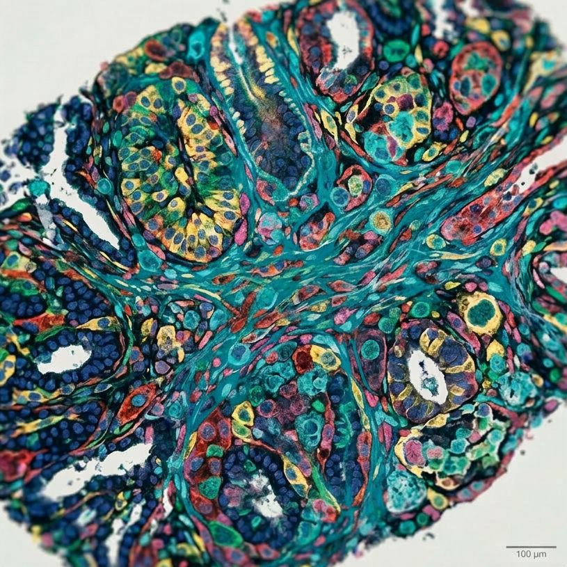
Biological tissues are inherently heterogeneous. Tumours in particular are composed of phenotypically and functionally distinct cell populations — malignant cells, stromal fibroblasts, immune infiltrates, and endothelial cells — whose individual proteomic states cannot be resolved by bulk approaches which averages signals across millions of cells. Deep visual proteomics directly addresses this challenge by enabling morphology-guided, spatially resolved proteomic profiling.
For oncology, deep visual proteomics illustrates how different cellular compartments within a tumour harbour distinct protein expression programmes, providing mechanistic insight into treatment resistance, disease progression, and microenvironmental crosstalk. In the context of diagnostics and precision medicine, this capability enables the identification of cell-type-specific protein biomarkers that are invisible to conventional bulk proteomics, opening pathways towards more accurate patient stratification and the development of targeted therapeutic strategies.
What is the Key Challenge of Deep Visual Proteomics?
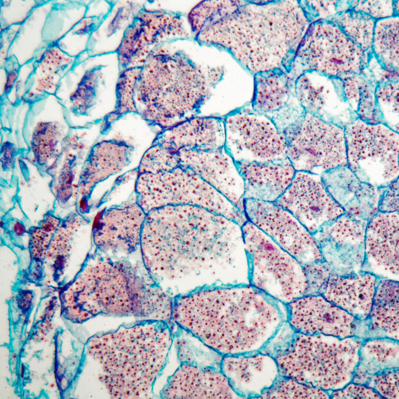
A major challenge in deep visual proteomics is the precise isolation of small regions of interest (ROIs). Single mammalian cells measure approximately 10–20 µm in diameter, whilst subcellular compartments, such as nuclei or specific organelles, can be smaller than 5 µm. Capturing these ROIs via laser microdissection (LMD) without compromising their integrity, while simultaneously sustaining high throughput, is challenging. This requires a sophisticated, gentle LMD platform that guarantees high efficiency and minimal sample loss, which can be further integrated with rapid, high-throughput processing in well-plate formats.
Integration of MMI CellCut into the Deep Visual Proteomics Workflow
Precision ROI Isolation
The MMI CellCut LMD system enables seamless integration into the deep visual proteomics workflow. As the only LMD system capable of precisely isolating complex ROI smaller than 10 µm, it sets a new standard for precision. Its short-pulse laser produces an exceptionally thin cutting edge of just 0.3 µm (using 5–10 µm FFPE tissue), enabling unmatched accuracy.
Isolate ROIs <5µm
High-Throughput Automated Collection
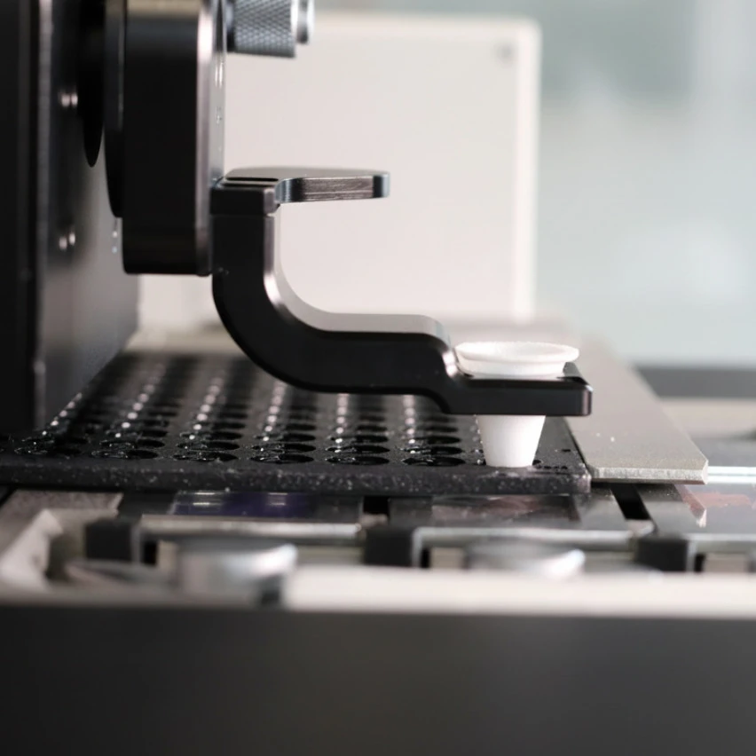
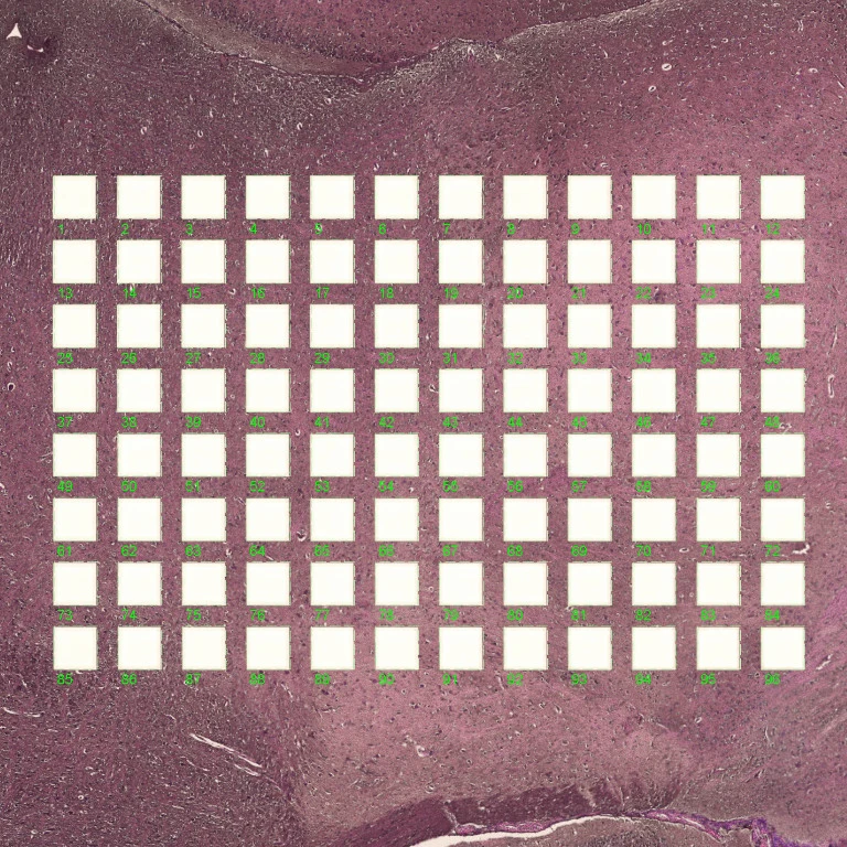
The MMI CellCut HT configuration supports fully automated collection into 96-well plates, enabling scalable, unattended processing of large sample cohorts. The system ensures that the maximum possible proteomic material from each ROI is recovered for deep visual proteomics analysis.
The image shows that all 96 shapes (liver FFPE, 5 µm) were successfully isolated using the CellCut HT.
95%+ Collection Efficiency
Cell Segmentation with Precise Alignment
Cell segmentation data in all standard formats—such as JSON, XML, CSV, TIFF, and HALO—can be imported directly and perfectly aligned with the live image, enabling more seamless workflows. Real-time verification confirms successful isolation after each dissection.
Supports all data formats
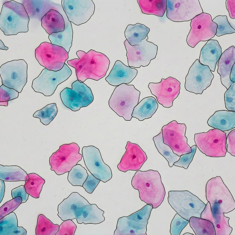
Isolation of a Cell Nucleus
using CellCut
Automated Laser Microdissection for Proteome Analysis
01
Sample Preparation
Patient tissue samples sectioned and prepared on LMD slides

02
High-Resolution Imaging
High resolution, whole-slide microscopy

03
AI-driven Image Analysis
Third-party image analysis AI software performs segmentation. Machine learning-based identification of cell types and states. Provision of ROIs for LMD

04
Automated Single Cell Laser Microdissection
Import ROI into LMD Software. Dissect directly into single tubes or 96-well plates. Isolate 96 single cells in less than 10 minutes
05
Ultra-High Sensitivity Mass Spectrometry
Liquid chromatography mass spectrometry (LC-MS)

06
Bioinformatic Data Analysis
Insights into the proteome in health and diseases at the cellular level
Revolutionising Pathology:
Why Mark Kühnel Trusts the MMI CellCut