

APPLICATIONS
Spatial Proteomics
Understanding Biology in Context
What is Spatial Proteomics?
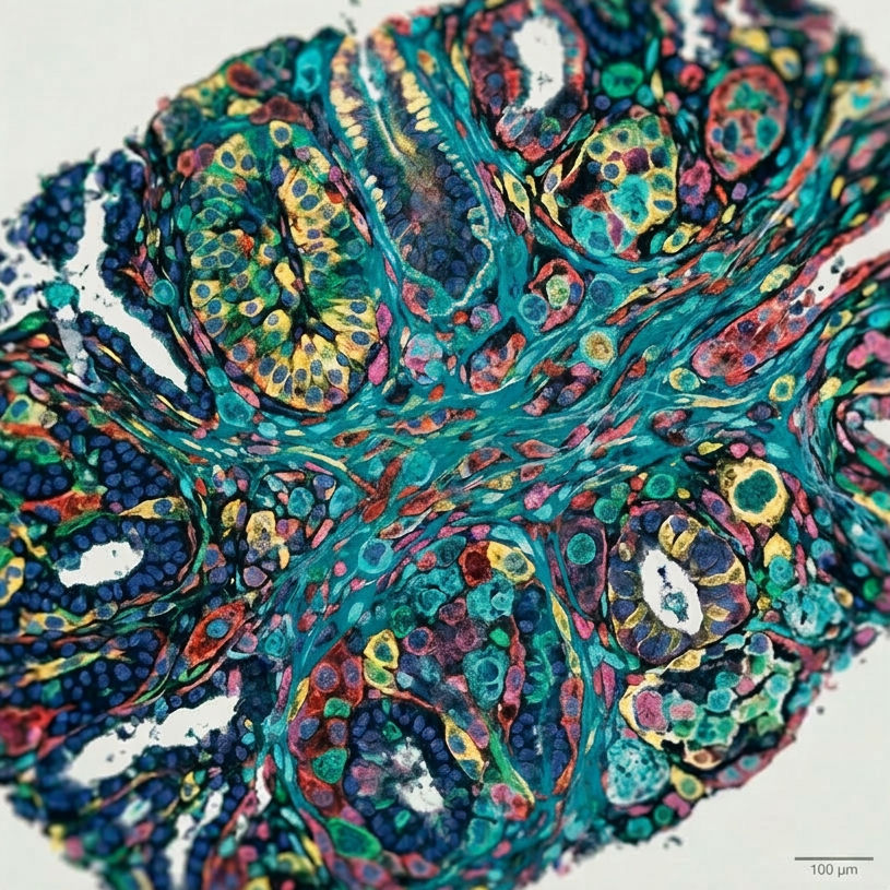
Spatial proteomics is an advanced field of proteomics that focuses on identifying and quantifying proteins while preserving their spatial distribution within cells and tissues. Spatial proteomics enables researchers to analyse proteins directly in their native biological environment, making spatial proteomics a powerful approach for understanding how protein localisation can influence function.
Unlike conventional proteomics methods, spatial proteomics retains positional information, allowing scientists to determine not only which proteins are present but also where they are located. This spatial dimension is essential, as spatial proteomics reveals how protein localisation within specific cellular compartments and tissue regions drive biological processes.
Why Spatial Proteomics Matters
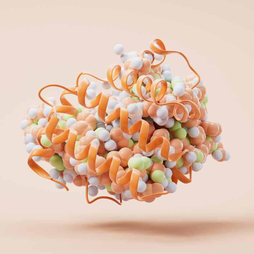
Spatial proteomics plays a critical role in advancing modern biology because spatial proteomics captures the organisation of proteins within cells and tissues. Many biological processes depend on precise protein localisation, and spatial proteomics allows researchers to investigate these processes with high resolution and precision.
In disease research, spatial proteomics is particularly important. Spatial proteomics enables the analysis of heterogeneous tissues, such as tumors, where different regions exhibit distinct molecular characteristics. By applying spatial proteomics, researchers can identify spatially resolved biomarkers, understand disease progression, and uncover new therapeutic targets.
Deep Visual Proteomics
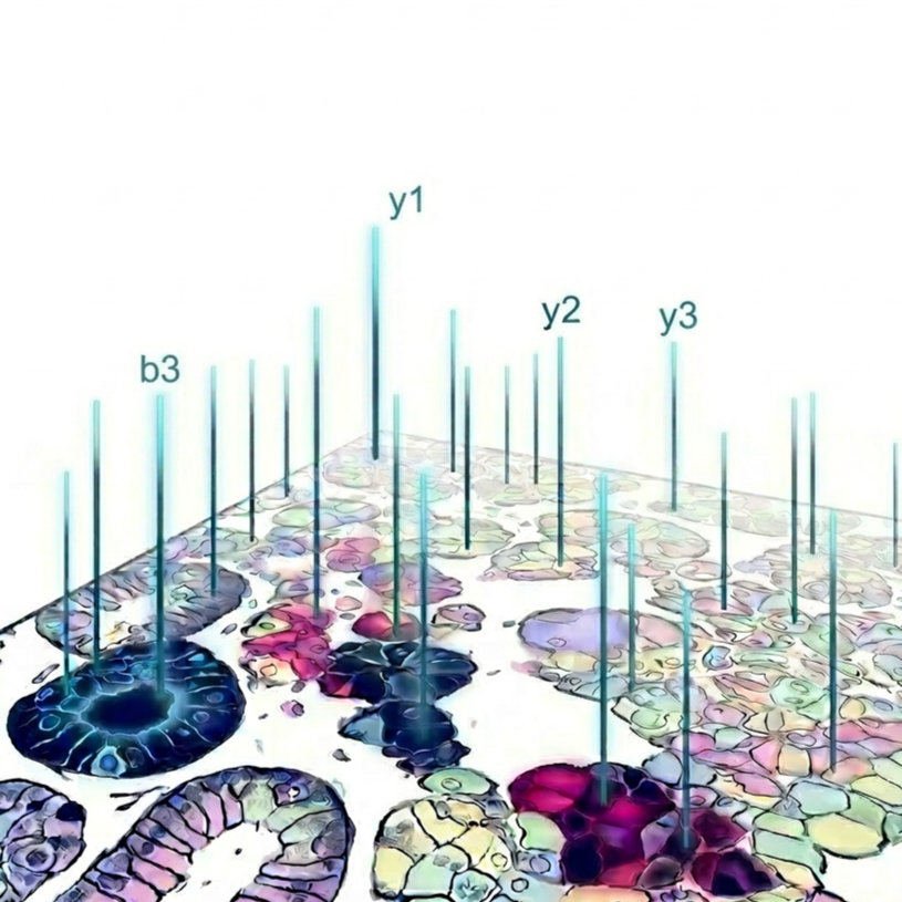
Deep visual proteomics (DVP) represents a next-generation approach within spatial proteomics that combines artificial intelligence, imaging, and proteomic analysis. DVP extends the capabilities of spatial proteomics by linking visual phenotypes with proteomic data at single-cell resolution.
Within spatial proteomics workflows, DVP uses machine learning to identify specific cells based on their morphology. These cells are then isolated using laser microdissection and analysed by mass spectrometry. This makes deep visual proteomics a powerful extension of spatial proteomics, enabling direct connections between tissue structure and molecular information.
Advanced Spatial Proteomics with MMI CellCut
Laser microdissection (LMD) is a key technology for spatial proteomics, enabling precise isolation of single cells or larger regions of interest (ROIs) while preserving spatial context. This is essential for linking protein localisation for histology and is particularly important for deep visual proteomics, where selected cells must be isolated with high accuracy for downstream analysis.
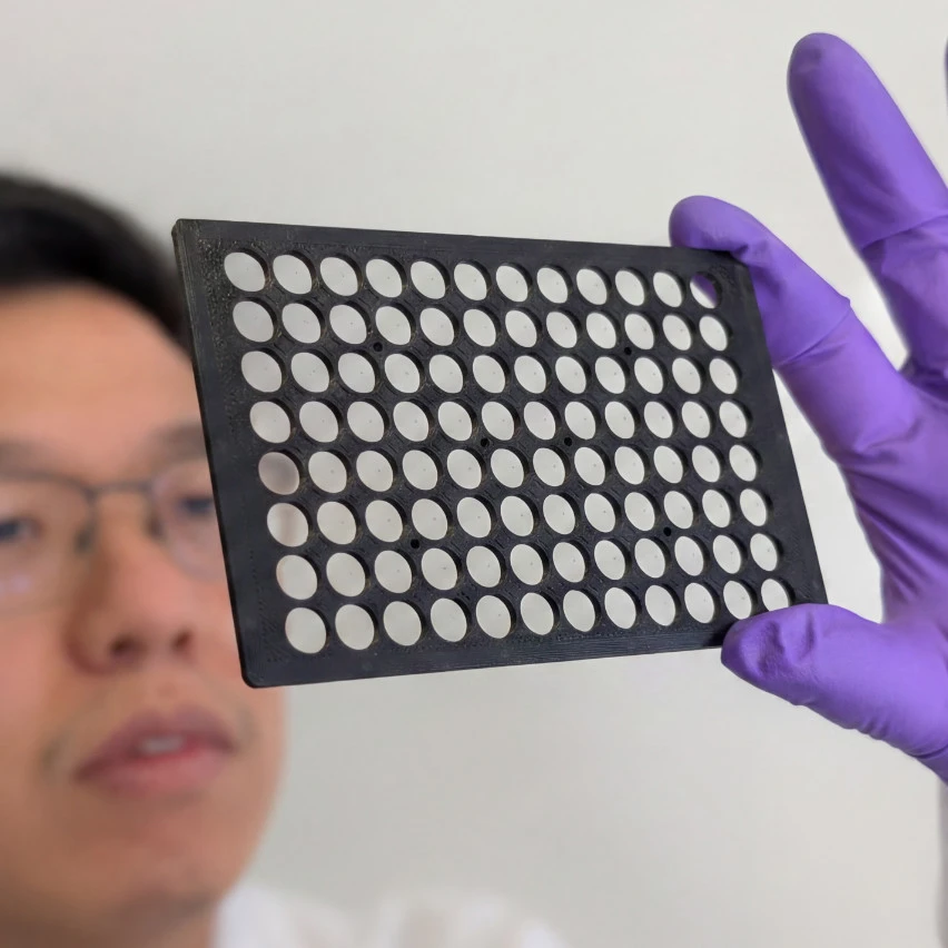
MMI provides advanced solutions for spatial proteomics with the MMI CellCut system, enabling efficient and highly precise isolation of single cells as well as subcellular compartments. The ROIs can be identified using separate AI-based cell segmentation software and then imported and aligned into the LMD system, enabling automated and reproducible workflows. For high-throughput applications, cells can be directly isolated into 96-well plates using the CellCut HT system.
95%+ Collection Efficiency
Automated Workflow for Spatial Proteomics
The MMI CellCut system integrates seamlessly into automated spatial proteomics workflows.

01
Sample Preparation
Preparation of the sample on Membrane Slides

02
Whole Slide Imaging
Whole-slide high-resolution microscopy to gain overview of the sample.

03
Cell Segmentation
Image analysis software performs image segmentation using deep learning to identify ROIs.

04
Automated LMD
Import cell shapes (ROIs) into LMD software. Dissect hundreds of single cells into reaction tubes or 96-well plates.

05
Mass Spectrometry
Gain cellular insights into the proteome
Isolation of a Cell Nucleus using CellCut
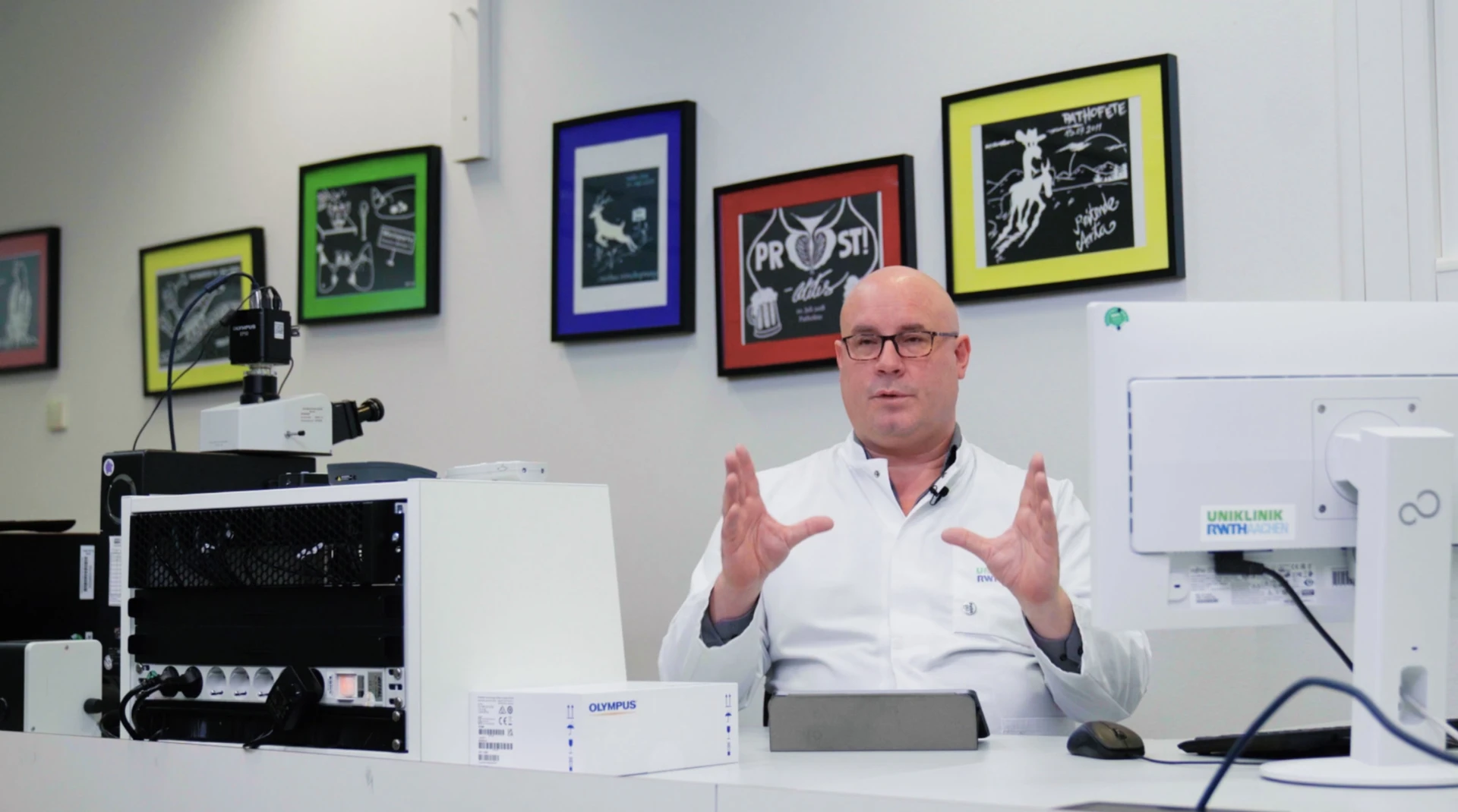
Revolutionising Pathology:
Why Mark Kühnel Trusts the MMI CellCut